Case Report
Surgical Resection of Cardiac Rhabdomyoma in a Neonate
Lugones Ignacio1,2*, Junco Nalá2 and Inguanzo Pablo Damián1
1Department of Pediatric Cardiology, Fundación FavaloroUniversity Hospital, Argentina
2Department of Pediatric Cardiology, Children´s Hospital of Buenos Aires “P. de Elizalde”, Argentina
*Corresponding author: Lugones Ignacio, Fundación FavaloroUniversity Hospital, Buenos Aires, Argentina, Av Manuel Montes de Oca 40, C1270AAN, Buenos Aires, Argentina
Published: 31 Aug, 2016
Cite this article as: Ignacio L, Nalá J, Damián IP. Surgical
Resection of Cardiac Rhabdomyoma in
a Neonate. Ann Clin Case Rep. 2016;
1: 1115.
Abstract
Rhabdomyomas are the most common cardiac tumors in infancy and childhood. Surgery is reserved for those patients with hemodynamic compromise. We present a case of surgical resection of a large mass obstructing the left ventricular outflow tract in a neonate.
Keywords
Rhabdomyoma; Cardiac tumor; Left ventricular outflow tract; Neonate
Case Presentation
A 38-weeks gestational age newborn girl was derived to our institution with the diagnosis of
a cardiac tumor. On her first day of life, a cardiac murmur and signs of low cardiac output were
detected. The echocardiogram performed after birth revealed the presence of a large mass in the left
ventricular outflow tract. Clinical presentation resembled that of critical aortic stenosis. Therefore,
prostaglandin infusion was started to restore adequate systemic blood flow and perfusion of vital
organs. The patient was derived to our institution and arrived in good condition. The diagnosis was
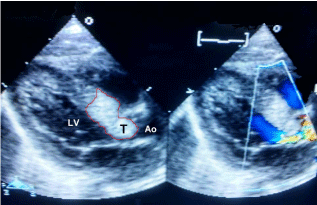
confirmed. The large tumor (19 x 17 mm) infiltrated the ventricular septum and protruded into
the left ventricular outflow tract, occluding 90% of its diameter (Figure 1). No other cardiac masses
were found.
All other organs were scanned for associated lesions using ultrasound and MRI. No other tumor
was found.
After diagnostic confirmation, the patient was taken to the operating room. Median sternotomy
was performed. Cardiopulmonary bypass was started using aortic and single venous cannulation.
The aorta was crossclamped. The ascending aorta was transected some millimeters above the
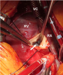
sinotubular junction. Through the aortic valve, the left ventricular outflow tract was explored. A
large homogeneous white mass was found just below the leaflets of the aortic valve. It completely
occluded the subaortic region, making visualization of the ventricular cavity impossible (Figure 2).
After identifying its free margins, the cardiac mass was detached from the ventricular septum
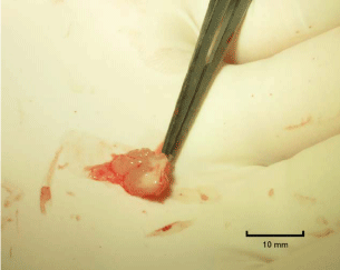
using an ophthalmic micro scalpel. The protruding portion was completely resected (Figure 3), but
part of the tumor remained infiltrating the ventricular septum. Extended resection in that area was
considered impossible without causing severe damage to vital anatomic structures. The aorta was
then sutured. Air was removed and the aortic cross clamp was released. The heart showed adequate
contractility with sinus rhythm. Cardiopulmonary bypass was ended and the chest was closed.
The patient was extubated on the following day, and discharged 5
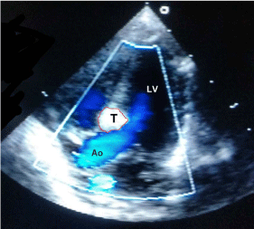
days later in an excellent condition. Postoperative echocardiogram
showed unobstructed left ventricular outflow tract without aortic
regurgitation or stenosis (Figure 4).
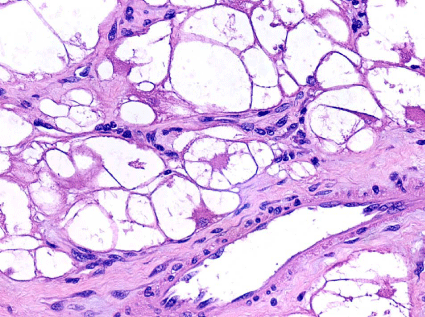
Anatomic pathology confirmed the diagnosis of rhabdomyoma
(Figure 5). Although there were no obvious signs of tuberous sclerosis,
the patient was derived to the geneticist after hospital discharge.
Figure 1
Figure 1
Long paraesternal view of the left ventricular outflow tract. The tumor infiltrates the ventricular septum
and protrudes into the subaortic region, leaving only a narrow pathway. The red line delimitates the margins of
the cardiac mass (Ao: Aorta; LV: Left Ventricle; T: Tumor).
Figure 2
Figure 2
The aorta has been transected above the sinotubular junction.
Through the aortic valve, a white mass can be identified occupying the left
ventricular outflow tract in almost all its diameter (AAo: Ascending Aorta; RA:
Right Atrium; RV: Right Ventricle; VC: Venous Cannula; Arrow; Tumor).
Figure 3
Figure 4
Figure 4
Five chamber apical view of the subaortic area showing
unobstructed pathway from the left ventricle to the ascending aorta. The size
of the tumor has been considerably reduced (Ao: Aorta; LV: Left Ventricle;
T: Tumor).
Figure 5
Discussion
Rhabdomyoma is the most prevalent of all cardiac tumors in
infancy and childhood [1]. It accounts for 35% of the cases. More
than 70% of the patients present with symptoms. It is a benign tumor
with a clear tendency to spontaneous regression and unreported
malignant degeneration. However, it can be life-threatening in those
cases in which it causes severe mechanical obstruction within the
heart. Left ventricular outflow tract obstruction is the most frequent
cause of death, and it is usually associated to a single tumor located
in the septum [2].
Surgical resection is reserved for these situations or to prevent
potentially lethal acute complications such as embolization or
ventricular arrhythmias [3]. Reported mortality rate is less than 5%. Previous studies have demonstrated no difference between total and
partial resection of cardiac rhabdomyomas in terms of postoperative
complications and mortality [4]. Besides, regrowth of the tumor after
partial excision is not common. Therefore, resecting only those parts
that produce hemodynamic alterations may be the safest strategy [3].
Masses with subaortic location are especially challenging, because
surgical approach must be done through the aortic valve, which has a
small diameter in the neonatal period [5].
This tumor is strongly related to tuberous sclerosis. Depending
on the series, 30% to 90% of patients with a cardiac rhabdomyoma
suffer from this disease. This is a genetic disorder with variable
compromise caused by benign tumors in many vital organs such
as the heart, central nervous system, eyes, kidneys, lungs and skin.
Prognosis is extremely variable and depends on the severity of the
manifestations [6].
References
- Burke A, Virmani R. Tumors of the heart and great vessels. Atlas of tumor pathology. 3rd series, AFIP, Washington DC. 1996.
- Verhaaren HA, Vanakker O, De Wolf D, Suys B, Francois K, Matthys D. Left ventricular outflow obstruction in rhabdomyoma of infancy: metaanalysis of the literature. J Pediatr. 2003; 143: 258-263.
- Padalino MA, Vida VL, Boccuzzo G, Tonello M, Sarris GE, Berggren H, et al. Surgery for primary cardiac tumors in children: early and late results in a multicenter European Congenital Heart Surgeons Association study. Circulation. 2012; 126: 22-30.
- Jacobs JP, Konstantakos AK, Holland FW, Herskowitz K, Ferrer PL, Perryman RA. Surgical treatment for cardiac rhabdomyomas in children. Ann Thorac Surg. 1994; 58: 1552–1555.
- Black MD, Kadletz M, Smallhorn JF, Freedom RM. Cardiac rhabdomyomas and obstructive left heart disease: histologically but not functionally benign. Ann Thorac Surg. 1998; 65: 1388-1390.
- Bosi G, Lintermans JP, Pellegrino PA, Svaluto-Moreolo G, Vliers A. The natural history of cardiac rhabdomyoma with and without tuberous sclerosis. Acta Paediatr. 1996; 85: 928-931.

