Case Report
Lobulation of the Testicles: A Case Report
Mahmoud Elnagar1* and Ravi Kadasne2
1Elnagar Urology Center, Egypt
2Emirates International Hospital, United Arab Emirates
*Corresponding author: Mahmoud Elnagar, Elnagar Urology Center, 35631, Belqas, Egypt
Published: 24 Aug 2016
Cite this article as: Elnagar M, Kadasne R. Lobulation of
the Testicles: A Case Report. Ann Clin
Case Rep. 2016; 1: 1094.
Abstract
Testicular lobulation is a rare congenital anomaly of the testicle. Scrotal ultrasound as a primary
diagnostic modality of testicular abnormalities is very helpful in the diagnosis of testicular lobulation.
We are reporting in bilateral testicular lobulation associated with varicocele and primary infertility.
The m=lobulation was discovered accidentally during routine scrotal ultrasound.
Keywords: Testicular lobulation; Scrotal ultrasound; Infertility; Congenital anomaly
Case Presentation
We are reporting on a rare testicular anomaly which is benign testicular lobulation in a male
with primary infertility.
A-30-year old male presented to urology clinic complaining of inability to conceive after two
years of marital relationship. The patient is neither diabetic nor hypertensive and does not receive
any medication for any chronic illness. The patient denied any history of surgical operation or
trauma. On local clinical examination of the scrotum, there was palpable, and visible varicocele
on the left side and the surface of the left testicle was irregular with palpable notch. Right testicle
was felt normal. Semen analysis revealed moderate asthenospermia with normal shape sperms.
Testicular grey scale and Doppler ultrasound showed normal size and architecture of both testicles
and dilated veins of the left spermatic cord with regurgitation of blood during Valsalva maneuver.
There were a deep longitudinal cleft on the surface of the left testicle (Figure 1) which appeared complete and a similar cleft on the surface of the right testicle but appeared incomplete (Figure 2). However, 3-D ultrasound could
show that the clefts were not complete on either side, and there was
bilateral testicular lobulation (Figure 3 and 4). The primary diagnosis
was settled as primary infertility due to poor sperm quality related to
the presence of grade III left varicocele.
Figure 1
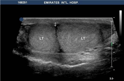
Figure 1
Grey scale ultrasound of the left testicle with deep longitudinal cleft (arrow) on the surface which
appears complete.
Figure 2
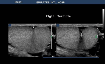
Figure 2
Grey scale ultrasound of the right testicle with a deep longitudinal cleft on the surface (arrow).
Figure 3
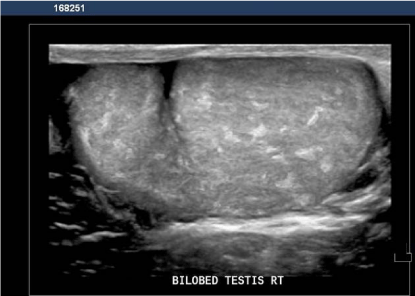
Figure 3
3-D ultrasound showed incomplete lobulation of the right testicle.
Discussion
Testicular lobulation is a rare anomaly of the testicles and has been reported [1]. This lobulation may be an idiopathic embryologic variant. The exact aetiology is unknown but is thought be a form of incomplete polyorchidism. It has been proposed that bilobed testis results from incomplete division of the urogenital ridge [2,3]. However, it has been suggested that testicular lobulation could be a sequence of orchiopexy and fibrosis after orchiopexy could be the reason for testicular lobulation [4,5]. In our case, the lobulation is bilateral, and the patient has no history of orchiopexy or inguinal surgery. Irregular testicular contour during physical examination can be differentiated from testicular malignancy by using ultrasound. However, careful ultrasound examination in different planes is essential for establishing correct diagnosis especially if the septum is incomplete. Moreover, 3-D ultrasound could be helpful in identification of the depth of the cleft. Testicular lobulation should be differentiated from polyorchidism and supernumerary testis. Polyorchidism is defined as the presence of more than two testes within the scrotum [6]. In most cases of polyorchidism, the extra testis is located on left side, and there are two epididymides and a single vas deferens [7]. In testicular lobulation there is a single epididymis with its vas as could be identified in our case and the lobulation is incomplete. While testicular lobulation is a benign condition the neoplasm of the supernumerary testis was found in 6.4% of cases [8].
Figure 4
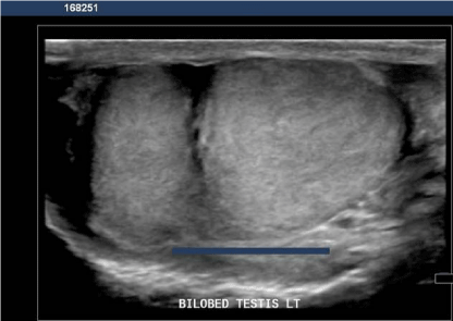
Figure 4
3-D ultrasound showed incomplete lobulation of the left testicle
References
- Kao EY, Gerscovich EO. Benign testicular lobulation: sonographic findings. J Ultrasound Med. 2003; 22: 299-301.
- McAlister W, Manley C. Bilobed testicle. Pediatr Radiol. 1987; 17: 82-82.
- Beiko D, Macneily AE. Torsion of bilobed testis and biopsy-proven ipsilateral supernumerary testis in an adolescent. Can Urol Assoc J. 2013; 4: E67-E70.
- Mihmanli I, Kantarci F. Benign testicular lobulation [letter]. J. Ultrasound Med. 2003; 22: 1001-1002.
- Kantraci F, Mihmanli I, Yilmaz MH, Cetinkaya S, Selcuk D, Ogut G. Orchiopexy: a cause of benign testicular lobulation. J. Ultrasound Med. 2003; 22: 1417-1419.
- Oner AY, Sahin C, Pocan S, Kizilkaya E. Polyorcidism: sonographic and magnetic resonance image findings. Acta Radiol. 2005; 46: 769-771.
- Deveci S, Aygun C, Agildere AM, Ozkardes H. Bilateral double testis: evaluation by magnetic resonance imaging. Int J Urol. 2004; 11: 813-815.
- Bergholz R, Wenke K. Polyorchidism: a met-analysis. J Urol. 2009; 182: 2422-2427.