Case Report
Isolated Celiac Artery Injury Following Blunt Abdominal Trauma
Maghraby NHM* and Nemeth J
Emergency Medicine / Trauma & Disaster Management, McGill University, Canada
*Corresponding author: Nisreen Hamza M Maghraby, Emergency Medicine / Trauma & Disaster Management, McGill University, Montreal, 1001 Boulevard Decarie, Room CS16237.1, H4A 3J1, Montreal, Canada
Published: 21 Aug 2016
Cite this article as: Maghraby NHM, Nemeth J. Isolated Celiac Artery Injury Following Blunt Abdominal Trau ma. Ann Clin Case Rep. 2016; 1: 1089.
Abstract
Isolated celiac artery injury is a rare event, especially post blunt traumatic injury. In the event of
hemodynamically instability, surgical exploration and management is the obvious approach. There
is however no clear consensus on the management of hemodynamically stable patients with such
injury. We present a case of an isolated celiac artery injury which was managed conservatively
medically.
Keywords: Celiac artery injury; Blunt trauma; Visceral vascular injury; Conservative
management
Introduction
Vascular injuries represent approximately 3% of all trauma patients’ injuries and involve mainly
the extremities [1]. Although the true incidence of visceral vascular injury is unknown, the incidence
of celiac injuries is thought to be the least out of all visceral injuries, representing approximately 1%
[2].
The celiac artery or celiac trunk is the first artery that arises directly from the abdominal aorta,
measuring approximately 1.5cm in length, having three main branches: i) the left gastric artery,
ii) the common hepatic artery and iii) the splenic artery, supplying mainly the vital organs of the
foregut. Given the vital structures its branches supply, injury to the artery results in significant
mortality (as high as 75%), due mainly to hemorrhage and delayed visceral ischemia [3,4].
*Patient consent has been obtained to publish this case.
Case Presentation
A 39 year old male was brought to our trauma center by emergency medical services (EMS), post
frontal impact motor vehicle crash (MVC) at approximately 50km/hr. The patient was seat belted
and the airbags deployed. On arrival the patient was asymptomatic, vital signs recorded were normal
(Blood pressure 137/75 mm Hg, Pulse 86 bpm, Respiratory rate 16, O2 sat 97% Room air) and the
Glasgow Coma Score (GCS) was 15. In particular the abdominal exam was unremarkable and the
FAST (Focused Assessment with Sonography in Trauma) was negative. The results of laboratory
investigations were also within normal limits (VBG: pH 7.39, pCO2 49mm Hg, HCO3 28.7 mm/L,
Base excess 4 mm/L, Lactate 1.8 mm/L, CBC: Hgb 148 g/L, Hct 0.44 L/L, Platelets 264 10^9/L,
Chemistry: Creatinine 97 umol/L, INR 1.14, Liver profile: T.Bili 10umol/L, ALA 30u/L, ALP 76u/L).
Following evaluation by the trauma team, the patient underwent a total head/neck/torso CT
scan due to the high risk mechanism of injury. The aforementioned imaging revealed an isolated
celiac artery dissection (Figure 1A and B).
The vascular surgery consultation service was asked to evaluate the patient. Since the patient
was hemodynamically stable and not actively bleeding, the recommendations were to manage the
patient conservatively, to repeat a CT angiogram in 48hrs and to start aspirin.
The patient was admitted to the intensive care unit for the first 24hrs, and then discharged to the
ward for further observation. The patient remained asymptomatic during his hospital stay. A repeat
CT angiography revealed a stable injury with all 3 branches of the celiac truck originating from
the true lumen and patent (Figure 2). The patient was discharged home with outpatient vascular follow-up. He was seen 3 weeks later at vascular clinic, no further imaging was required. Patient
didn’t show up for his 6 months follow up, but has been following up regularly with his cardiologist.
Figure 1a
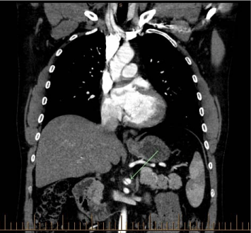
Figure 1a
Celiac Artery Dissection, injury initially identified on CT angio of
the chest.
Figure 1b
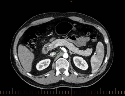
Figure 1b
Celiac Artery Dissection identified on CT abdomen (Trauma
protocol).
Discussion
Celiac artery injury results mainly (90-95%) from penetrating
injury, blunt trauma representing only 5-10% of cases [5]. Blunt
trauma results in celiac injury either directly from compression
against a bony structure (seat belt injury) or indirectly from the
deceleration forces especially in high speed collisions.
Diagnosis of such injuries also can present challenges if index of
suspicion is not high. Many trauma CT abdomen protocols capture
the images a few seconds to minutes after injection of the contrast
material, to obtain the portal phase of the imaging to improve views
of potential solid organ injury. This process however risks missing
the proper imaging of the arterial system [6]. As with our case,
the injury was incidentally identified in the lower cuts of the chest
CT angiography. This potential shortcoming of many trauma CT
abdomen protocols reemphasises the importance of repeating a CT
angiography of the abdomen in patients with abdominal trauma with
persistent abdominal symptomatology, to prevent detrimental delays
leading to potential detrimental clinical outcome.
In patients with hemodynamic instability, surgical exploration
and management is the obvious option. In hemodynamically stable,
asymptomatic patients however, the treatment options are unclear.
Most of the reported cases included symptomatic patients and were
managed either surgically or with endovascular methods [7].
Figure 2
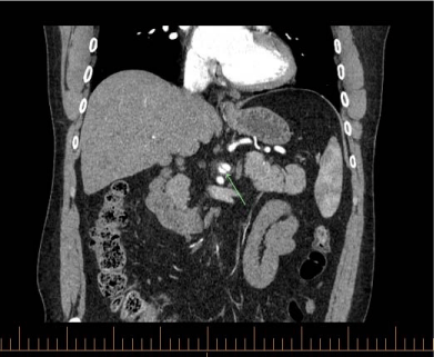
Figure 2
Follow up image 48hrs post injury with stable Celiac Artery
Dissection.
Conclision
Celiac artery injury is a rare with a high mortality rate if the
diagnosis is delayed. In hemodynamically unstable patients, surgical
treatment is required. In hemodynamically stable, asymptomatic
patients, the treatment options (surgical vs. endovascular repair
vs. conservative medical), the timing of reimaging and duration of
observation and are not as well defined.
A high clinical suspicion and low threshold for repetition of CT
imaging should be maintained for all trauma patients with persistent/
new abdominal symptoms. The differential diagnosis should include
arterial injury.
References
- Sirinek KR, Gaskill HV, Root HD, Levine BA. Truncal vascular injury factors affecting survival. J Trauma. 1983; 23: 372–377.
- Chlodwig K, Julia Stegmaier, Michael Krotz, Elisabeth Muetzel, Wolf Mutschler, Karl-Georg Kanz, et al. Celiac Dissection after Blunt Abdominal Trauma Complicated by Acute Hepatic Failure: Case Report and Review of Literature. Journal of Vascular Surgery. 2007; 576-580.
- Chaillou P, Moussu P, Noel SF. Spontaneous dissection of the celiac artery. Ann VascSurg. 1997; 11: 413–415.
- Brown DB, Singh H, Atnip RG, Cardella JF, Waybill PN. Blunt traumatic injury to the superior mesenteric artery and celiac axis. J Vasc Interv Radiol. 1998; 9: 783-785.
- Davis TP, Feliciano DV, Rozycki GS, Bush JB, Ingram WL, Salomone JP, et al. Results with abdominal vascular trauma in the modern era. Am Surg. 2001; 67: 565-570.
- Linsenmaier U, Krotz M, Hauser H, Rock C, Rieger J, Bohndorf K, et al. Whole-body computed tomography in polytrauma: techniques and management. EurRadiol. 2002; 12: 1728-1740.
- In Young Choi, Hwan Hoon Chung, Seung Hwa Lee, Sung Bum Cho, Yun Hwan Kim, Bo Kyoung Seo, et al. Treatment of a Traumatic Celiac Trunk Detachment by Bridging with a Stent Graft. CardioVascular andInterventional Radiology. 2012; 35: 422-425.