Case Report
Solid Pseudopapillary Neoplasm of the Pancreas
Yi-Wen Liao1, Vincent Chong1, Neil Wylie1, Neville Angelo2 and Jonathan Koea1*
1Department of Surgery, North Shore Hospital, New Zealand
2Department Pathology North Shore Hospital, New Zealand
*Corresponding author: Jonathan Koea, Department of Surgery, North Shore Hospital, New Zealand
Published: 18 Aug, 2016
Cite this article as: Liao Y-W, Chong V, Wylie N, Angelo
N, Koea J. Solid Pseudopapillary
Neoplasm of the Pancreas. Ann Clin
Case Rep. 2016; 1: 1084.
Abstract
A 35 year old presented with a 6 month history of an epigastric mass and upper abdominal
discomfort. Clinical examination confirmed a firm mass in the epigastrium but no signs of metastatic
disease. Investigations showed that tumour markers (carcinoembryonic antigen, Ca 19-9, Ca 125,
chromogranin A) were within the normal limits. A CT scan of the abdomen showed a large wellencapsulated
complex cystic mass with internal calcification, measuring 16 x 17 x 11.6 cm arising
in the tail of pancreas with a small focus of calcification. Following multidisciplinary review she
proceeded to a distal pancreatectomy and a splenectomy with the operative findings of a firm mass
arising in the pancreatic tail involving the splenic artery and vein. She was discharged on day 6 and
remains well 18 months post-operation.
Histological and immunohistochemical examination showed features consistent with solid
pseudopapillary neoplasm, with areas of necrosis and haemorrhage with tumour cells arranged
as discohesive nests and nuclear and cytoplasmic staining for vimentin and B-catenin. Solid
pseudopapillary neoplasm is a rare primary pancreatic tumour that usually presents in young
women with an abdominal mass and, while the primary tumours are often large, resection is usually
feasible and long term survival expected.
Keywords: Solid pseudopapillary neoplasm; Pancreatectomy
Introduction
Solid tumours of the exocrine pancreas are usually characterised by aggressive biology with local invasion and metastatic disease common findings at presentation making effective treatment difficult and resulting in poor survival [1,2]. Solid pseudopapillary neoplasm (SPN) is an unusual tumour that develops from the exocrine pancreas and tumours are often over 10 cm in diameter at presentation. However metastatic disease is rare and complete resection is usually possible with excellent long term survival [3].
Case Presentation
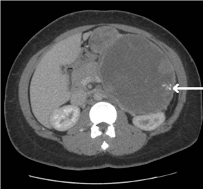
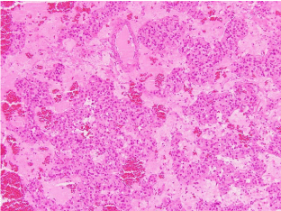
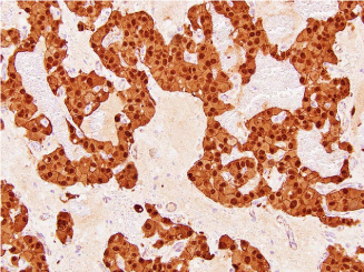
A 35 year-old Samoan woman was referred with a six-month history of a painless epigastric mass and weight loss. She had no significant previous medical or surgical history and physical examination was unremarkable except for a firm mass in the epigastrium. Full blood count, biochemistry and tumour markers (carcinoembryonic antigen, Ca 19-9, Ca 125, chromogranin A) were all within the normal limits. A CT scan of the abdomen showed a large well- encapsulated complex cystic mass with internal calcification, measuring 16 x 17 x11.6 cm arising in the tail of pancreas (Figure 1). There were no signs of distant spread or regional lymphadenopathy. Following multidisciplinary review she proceeded to a distal pancreatectomy and a splenectomy with the operative findings of a firm mass arising in the pancreatic tail involving the splenic artery and vein. No metastatic disease was seen and her post-operative course was uncomplicated. Histological and immunohistochemical examination showed features consistent with SPN, with areas of necrosis and haemorrhage with tumour cells arranged as discohesive nests accompanied by small blood vessels imparting a papillary configuration (Figure 2). There was a nuclear and cytoplasmic staining for vimentin and B-catenin (Figure 3). The patient remains alive and well 18 months post-operation and under regular follow up.
Figure 1
Figure 1
Axial CT scan demonstrating a large, heterogeneous soft tissue mass arising from the pancreas and containing a small area of calcification (arrow).
Figure 2
Figure 2
High power microscopy showing areas of necrosis and haemorrhage with tumour cells arranged in a papillary configuration associated with small blood vessels (haematoxylllin and eosin, 500 x magnification).
Figure 3
Figure 3
Immunohistochemical staining for β-catenin demonstrating nuclear uptake (500 x magnification).
Discussion
Tumours of the exocrine pancreas include adenocarcinoma, acinar cell carcinoma, intraductal
papillary mucinous neoplasm and mucinous cystadenocarcinoma. They are generally biologically aggressive and associated with poor survival. Solid pseudopapillary neoplasm develops from the exocrine part of the pancreas [1] but is an indolent tumour. It was first described by Virginia Frantz [2] and historically it accounts for 1% of all the pancreatic neoplasms, 5% of the cystic neoplasms and presents most commonly in women in their 30s but can occur between the ages of 13 and 60 years [3]. The tumours are usually large at presentation and presenting symptoms are related to the presence of a mass and early satiety [3]. The diagnosis is confirmed with typical imaging features demonstrating large encapsulated tumours with solid and cystic spaces [4]. Although pseudopapillary neoplasms are often large they are generally localised although between 9-15% may present with metastases or signs of local invasion [1-4]. Routine preoperative biopsy is not recommended as it is of limited utility and may result in dissemination [4]. SPN can occur throughout the pancreas but up to 60% develop in the tail or body. Treatment is surgical resection and the outcome following complete resection is excellent with overall 5 year survival of over 95% [5].
SPN are an increasingly important pancreatic lesion. A recent systematic review has shown that 90% of all cases have been reported in the 12 years and, of these 90% are now initially diagnosed as an incidental finding on cross-sectional imaging. Consistent with more early diagnosis is the observation that the mean size at presentation has decreased from 10 cm to 8 cm in the last decade [6]. These findings suggest that SPN is more common than initially thought and will become a significant part of the caseload for the pancreatic surgeon.
References
- Ren Z, Zhang P, Zhang X, Liu B. Solid pseudopapillary neoplasms of the oancreas: clinicopathologic features and surgical treatment of 19 cases. In J Clin Exp Pathol. 2014; 7: 6889-6897.
- Frantz VK. Tumors of the pancreas, Atlas of Tumor Pathology VII. In: Fascicles 27 and 28. Washington. Armed Forces Institute of Pathology, 1959; 47: 334.
- Cai Y, Ran X, Xie S, Wang X, Peng B, Mai G, Liu X. Surgical management and long-term follow-up of solid pseudopapillary tumor of the pancreas: A large series from a single institution. J Gastrointest Surg. 2014; 18: 935-940.
- Ganeshan DM, Paulson E, Tamm EP, Taggart MW, Balachandran A, Bhosale P. Solid pseudo-papillary tumours of the pancreas: current update. Abdom Imaging. 2013; 38: 1373-1382.
- Papavramidis T, Papavramidis S. Solid pseudopapillary tumors of the pancreas: review of 718 patients reported in English literature. J Am Coll Surg. 2005; 200: 965-972.
- Law JK, Ahmed A, Singh VK, Akshintala VS, Olson MT, Raman SP, et al. A systematic review of solid-pseudopapillary neoplasms: are these rare lesions? Pancreas. 2014; 43: 331-337.


