Case Report
Successful Management of Hematuria from Orthotopic Neobladder Varices with Transjugular Intrahepatic Portosystemic Shunt (TIPS) and Variceal Embolization
Justin Fincher1*, EhabEltahawy1 and Ruizong Li2
1Department of urology, University of Arkansas for Medical Sciences, USA
2Department of Interventional Radiology, University of Arkansas for Medical Sciences, USA
*Corresponding author: Justin Fincher, Urology Resident, University of Arkansas for Medical Sciences, 4301 W Markham St# 540, Little Rock 72205, AR, USA
Published: 12 Aug, 2016
Cite this article as: Fincher J, Eltahawy E, Li R. Successful
Management of Hematuria from
Orthotopic Neobladder Varices with
Transjugular Intrahepatic Portosystemic
Shunt (TIPS) and Variceal
Embolization. Ann Clin Case Rep.
2016; 1: 1064.
Abstract
Hematuria in an orthotopic neobladder is a difficult entity to manage due to its' unique anatomic and physiologic features. Infection, stone disease, primary bowel mass, and recurrent urothelial cell cancer are among the causes to consider. Here, we report a case of a 61 year old male who developed hematuria secondary to neobladder varices which was managed with TIPS (Transjugular Intrahepatic Portosystemic Shunt) and variceal embolization.
Keywords: Neobladder; Hematuria; TIPS; Cirrhosis; Bladder cancer; Varices
Introduction
The standard of care for muscle invasive bladder cancer is a radical cystectomy. While different urinary diversion techniques are utilized, an orthotopicneobladder allows the patient to utilize the natural orifice for urine drainage [1]. By utilizing bowel, neobladders are uniquely susceptible to pathologies associated with the gastrointestinal system as well as the genitourinary system [2]. This creates a diagnostic problem in these patients when presenting with hematuria. This is a case report of a male with hepatic cirrhosis who developed hematuria from neobladder varices that was managed by TIPS and variceal embolization. To our knowledge, this is the first case of TIPS and variceal embolization being utilized to successfully stop hematuria in this setting.
Case Presentation
A 61 year old male with idiopathic hepatic cirrhosis who had undergone cystoprostatectomy for muscle invasive bladder cancer with ileal neobladder ten years prior presented with a one month history of hematuria. He maintained follow up after his original surgery as it was complicated by wound dehissence resulting in neobladder cutaneous fistula. It was eventually closed and he was lost to follow up about 7 years ago. He reports that he was doing well in the interim until he developed hematuria but did not seek treatment until he developed clot retention and went to an outside hospital. There, he had cystoscopy which revealed diffuse mucosal hemorrhage. He continued to have hematuria requiring continuous bladder irrigation and blood transfusion so he was transferred to our academic institution. On presentation, he was actively hemorrhaging from his neobladder so he was admitted to the medical intensive care unit (ICU). His liver function was compromised as he had Child Pugh class B cirrhosis with total bilirubin of 1.5mg/dL, albumin at 2.4g/dL, prothrombin time of 16.7s, moderate ascites on CT scan, and no evidence of encephalopathy. After the patient received resuscitation, cystoscopy was performed to evacuate clots revealing hemorrhage emanating from multiple sites but no mucosal abnormalities indicative of neoplasm. CT angiogram did not show active extravasation but did show prominent varices surrounding the neobladder (Figure 1). During the diagnostic workup, he remained in the ICU as his hematuria persisted such that he received a total of 20 units of packed red blood cells, 6 units of platelets, and 8 units of fresh frozen plasma to maintain hemodynamic stability. The medical team’s recommendation was to obtain hepatic venous pressure gradient measurement to assess the varices as a cause of the bleeding. His pressure gradient was found to be elevated at 19 mmHg (normal <5mmHg) so 10 mm TIPS endoprosthesis consisting of a nitinol stent with expanded polytetrafluoroethylene (ePTFE) graft was placed lowering the gradient to 6 mmHg [3] (Figure 2 and 3). Unfortunately, variceal flow remained retrograde and his bleeding continued 2 days later so embolization of the neobladder varices was performed with vascular coils and Amplatzer plugs (Figure 4). Subsequently, his hematuria resolved. He remained free of signs of TIPS complications without development of encephalopathy, fever, or worsening ascites. He was able to void spontaneously by Crede, external compression of the bladder, after foley catheter removal and had low urine residual before discharge.
Figure 1
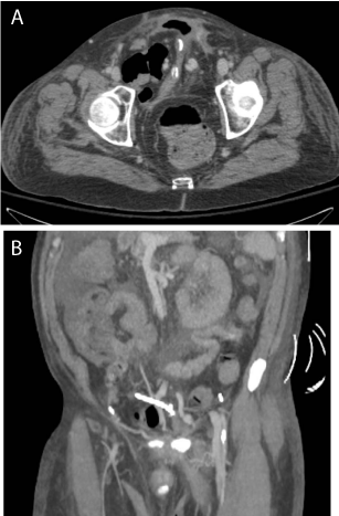
Figure 1
(A) Axial and (B) coronal abdomen CT images showing prominent varicose veins around the neobladder.
Figure 2
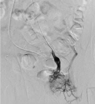
Figure 2
Menseteric venogram during TIPS procedure showing varicose veins and systemic shunt to the iliac veins.
Figure 3
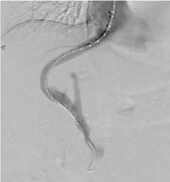
Figure 3
TIPS shunt between right hepatic vein and right portal vein.
Figure 4
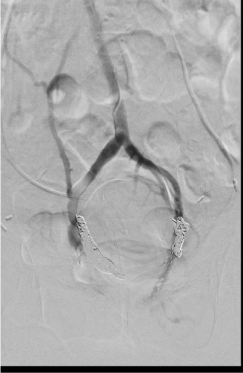
Figure 4
Post varicose veins embolization with amplatzer vascular plug and multiple nitinol coils.
Discussion
Hematuria as a result of neobladder varices is a rare entity. Review of the literature only revealed one other case. That patient was unsuccessfully treated with diathermy of the mucosa at the sites of hemorrhage resulting in his death [4]. Additionally, there is a handful of case reports of patients who developed hematuria from varices associated with an ileal conduit. Two of these were successfully treated by a TIPS procedure while another was managed by revision of the conduit and ligation of the varix [5-7].
Although very few cases have been reported, the etiology of the bleeding suggests this pathology carries a high risk of death. In the current case, the patient was improved a combination of TIPS and variceal embolization. Additionally, he benefitted from having relatively preserved liver function in the face of cirrhosis severe enough to induce varices and ascites.
Conclusion
The causes of hematuria in a patient with an orthotopic neobladder can range from easily treatable causes like infection or stone to life threatening causes like recurrent cancer or neobladder varices. The difficulty associated with managing patients who develop hematuria from varices mandates that liver function and cirrhotic state are factored in when considering type of urinary diversion. Based off the limited number of case reports available, ileal conduit may be a safer option in these patients as management of hematuria from the varices appears to be more successful. Although many factors influence the overall survival of a patient with muscle invasive bladder cancer considering radical cystectomy, underlying hepatic disease must be included. We present a treatment option for patients who develop hematuria as a result of portal hypertension induced neobladder varices.
References
- Hautmann RE, Abol-Enein H, Lee CT, Mansson W, Mills RD, Penson DF, et al. Urinary diversion: how experts divert. Urology. 2015; 85: 233-238.
- Hautmann RE, Volkmer BG, Schumacher MC, Gschwend JE, Studer UE. Long-term results of standard procedures in urology: the ilealneobladder. World J Urol. 2006; 24: 305-314.
- Garcia-Pagan JG, Groszmann R, Bosch J. Portal hypertension WM Weinstein, CJ Hawkey, J Bosch. Portal hypertension. 1st ed. Philadelphia:Elsevier Mosby; 200: 707–716.
- Oderda M, Mondino P, Lucca I, Fiorito C, Gillo A, Zitella A, et al. Fatal haematuria in a patient with an orthotopicneobladder and chronic liver failure. Urologia Internationalis. 2009; 83: 368-369.
- Dal Moro F. Long-delayed gross hematuria due to portal hypertension in an alcoholic cirrhotic patient with ileal conduit urinary diversion. Ann Hepatol. 2014; 13: 830-831.
- Felig DM, Bleecker DP, De Meritt DS. Hematuria in a patient with an ileum conduit and hepatic cirrhosis. N Engl J Med. 2001; 344: 939.
- Tu WH, Chao D, Gill H. Gross hematuria from an ileal conduit as a first presentation of portal hypertension. Nat ClinPract Urol. 2008; 5: 569-573.