Case Report
Soft Palate Schwannoma: A Rare Case of an Intraoral Mass
Shi LL1, Justicz N2, Panella NJ2 and Henriquez OA2*
1College of Pharmaceutical Sciences, Soochow University, People's Republic of China
2Department of Otolaryngology -Head and Neck Surgery, Emory University School of Medicine, USA
*Corresponding author: Henriquez OA, Department of Otolaryngology-Head and Neck Surgery, Emory University School of Medicine, 550 Peachtree St. NE Suite 1135, Atlanta, GA 30308, USA
Published: 05 Aug, 2016
Cite this article as: Shi LL, Justicz N, Panella NJ,
Henriquez OA. Soft Palate
Schwannoma: A Rare Case of an
Intraoral Mass. Ann Clin Case Rep.
2016; 1: 1057.
Abstract
Introduction: Schwannomas are benign encapsulated neoplasms that arise from proliferation of
Schwann cells and can originate from any cranial, peripheral, or autonomic nerve. While most
schwannomas present in the head and neck region, only 1% of these occur intra orally and these
typically occur on the tongue or floor of mouth.
Case: We report here a 55-year-old woman seen at our institution who presented with a mass of
the soft palate. Biopsy found it to be a schwannoma of the soft palate, which was surgically excised.
Conclusion: Because of the predilection of schwannomas to occur in the head and neck region,
otolaryngologists must maintain a high level of suspicion for unique and rare presentations.
Keywords: Schwannomas; Neoplasms; Glossotonsillar sulcus
Introduction
Schwannomas, first described by Verocay in 1910, are best characterized as benign, encapsulated neoplasms that arisefrom proliferation of Schwann cells within the perineurium. Hence, theycan originate from any cranial, peripheral, or autonomic nerve [1]. Their pathophysiology is thought to be secondary to loss of function of merlin, a cytoskeleton protein, either by primary alteration of the NF2 gene on chromosome 22 or by inactivation of the merlin protein [2]. The head and neck region is the most common site for schwanommas;according to Das Gupta et al. [3], 45% of a series of 303 benign nerve sheath tumors occurred within this locale. However, only 1% of head and neck schwannomas occur intra orally. They are most commonly in the mobile portion of the tongue or the floor of the mouth and less frequently in the palate [4,5]. Here, we present the case of a schwannoma of the soft palate seen at our institution. The patient provided written consent as case reports are IRB exempt at our institution.
Case Presentation
A 55-year-old African American female with a history of obstructive sleep apnea presented for
evaluation of a soft palate mass. During a dental procedure, a dome-shaped mass on the soft palate
was noted, and a resultant CT scan of the neck with contrast showed a 1.6 x 2 cm heterogeneous
soft tissue mass localized to the lateral oropharyngeal wall and glossotonsillar sulcus concerning
for intraoral malignancy. The patient had not noticed the mass previously. She denied a history of
smoking, excessive alcohol consumption, or radiation exposure. She reported no recent weight loss,
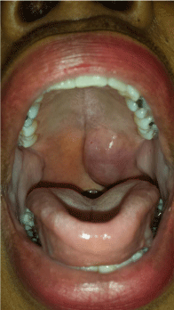
no change in her voice, no difficulty breathing, and no otalgia. The only significant finding on exam
was the submucosal, rubbery mass without mucosal changes or ulceration, which did not cross the
midline (Figure 1). It was non-tender, non-cystic, and non-fluctuant. Flexible laryngoscopy was
within normal limits.
A biopsy was taken and several small pieces of rubbery, yellow tissue were obtained. No fluid
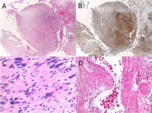
was expressed. Immunohistochemical studies revealed that the tumor cells were positive for S100
and negative for AE1/AE3 cytokeratin (Figure 2A and B). Permanent sectioning demonstrated
classic well-formed nuclear palisades surrounding the fibrillary processes (Figure 2C) as well as
large, irregular vessels with hyalinization and rare thrombi (Figure 2D). These findings were most
consistent with a schwannoma.
Surgical excision was performed under general anesthesia and exposure was obtained using a
McIvor mouth gag. The lesion dissected easily away from the posterior hard palate and the palatal
musculature. The specimen was 3.5 x 2.2 x 1.9 cm in size. The defect was closed primarily with a
combination of absorbable suture and a small piece of AlloDerm® (Acelity, San Antonio, TX) graft. At follow-up eleven days later, the surgical site was healing well and the AlloDerm® was no longer identifiable within the wound bed.
Figure 1
Figure 2
Figure 2
Hematoxylin-eosin (H&E), original magnification x40
(A) With biopsy specimen showing tumor cells stained positive for S-100
(B) H&E, original magnification x400
(C) Demonstrating well-formed nuclear palisades surrounding fibrillary processes, and H&Ex200
(D) Demonstrating large, irregular vessels with hyalinization and rare thrombi.
Results and Discussion
This case represents an unusual presentation of a schwannoma, identified on biopsy before definitive excision. The initial biopsy revealed a spindle cell neoplasm composed of long, thin cells with tapered nuclei, coarse chromatin, and ill-defined cytoplasm proliferating in a biphasic pattern. Focal areas suggestive of the presence of Verocay bodies were confirmed on the surgical specimen. Classically, Schwannomas have an Antoni A palisading hypercellular component with Verocay bodies and an Antoni B hypocellular and myxoid component.
Cytologically, the cells are elongated with hyperchromatic, wavy andtapered nuclei [2,5]. Immunohistochemical analysis (S-100, Leu-7) is often used to confirm and classify the diagnosis of nerve sheath tumors [5].
While this schwanomma was identified prior to surgery, diagnosis is not always possible until the tumor has been excised. Diagnosis and surgical planning can be aided by CT or MRI imaging to evaluate the extent of infiltration into surrounding tissues. The combination of imaging, biopsy, and immunochemical staining allows for appropriate diagnosis and surgical management. The definitive treatment of schwanomma is excision, and the causal nerve is often unlikely to be identified [5].
The differential diagnosis for a soft palate neoplasm is varied and includes minor salivary gland tumor, pyogenic granuloma squamous cell carcinoma, or lipoma [1]. Histologically, however, other neurogenic tumors such as neurofibroma, neuroma, myoblastoma of granular cells, neurogenic sarcoma, malignant schwannoma, neuroepithelioma and melanoma must also be excluded [5]. Schwannomas very rarely undergo malignant transformation [5].
Conclusion
While our original differential diagnosis included minor salivary gland tumors, torus palatinus, and squamous cell carcinoma, the histological findings allowed us to refine our diagnosis to a soft palate schwannoma. Schwannomas may present from any cranial, peripheral, or autonomic nerve. However, as nearly half of benign nerve sheath tumors occur in the head and neck, otolaryngologists must be particularly aware of the many unique presentations of schwannoma and maintain a high level of clinical suspicion. Early detection and biopsy can allow for appropriate surgical management. Schwannomas, therefore, should represent an important differential diagnosis for neoplasms in the head and neck.
Conclusion
The authors would like to acknowledge Dr. Shahrzad Ehdaivand from the Emory University Department of Pathology for her support obtaining the pathology slides pictures.
References
- Venkatachala S, Krishnakumar R, Rubby SA. Soft palate schwannoma. Indian J Surg. 2013; 75: 319-321.
- Hilton DA, Hanemann CO. Schwannomas and their pathogenesis. Brain Pathol. 2014; 24: 205-220.
- Das Gupta TK, Brasfield RD, Strong EW, Hajdu SI. Benign solitary Schwannomas (neurilemomas). Cancer. 1969; 24: 355-366.
- Shetty SR, Mishra C, Shetty P, Kaur A, Babu S. Palatal schwannoma in an elderly woman. Gerodontology. 2012; 29: e1133-1135.
- Colreavy MP, Lacy PD, Hughes J, Bouchier-Hayes D, Brennan P, O'Dwyer AJ, et al. Head and neck schwannomas-a 10 year review. J Laryngol Otol. 2000; 114: 119-124.


