Case Report
Low Grade Fibromyxoid Sarcoma of the Neck: A Case Report
Tatari MM*, Elhariti L, Abou-elfadl M, Abada RL, Rouadi S, Roubal M and Mahtar M
Department of Otorhinolaryngology, Africa
*Corresponding author: Tatari MM, Department of Otorhinolaryngology, face and Neck Surgery, 20 August 1953 Hospital, Casablanca, Morocco, Africa
Published: 04 Jul, 2016
Cite this article as: Tatari MM, Elhariti L, Abou-elfadl M,
Abada RL, Rouadi S, Roubal M, et al.
Low Grade Fibromyxoid Sarcoma of the
Neck: A Case Report. Ann Clin Case
Rep. 2016; 1: 1056.
Abstract
Low-grade fibromyxoid sarcoma (LGFMS) is a deceptively bland malignancy with potential for late recurrence and metastasis. It is a rare tumor but probably under-diagnosed because it is often confused with other entities. It is mainly localized in the deep soft tissues of proximal regions of the limbs and trunk. That makes this study very interesting for the neck localisation. So we descript the case of 70 years old women who came to our department with a big mass in the neck.
Keywords: Low grade fibromyxoid sarcoma; Head; Neck; Immunohistochemestry
Introduction
Low-grade fibromyxoid sarcoma (LGFMS) is a deceptively bland malignancy with potential for late recurrence and metastasis [1]. It is a rare tumor but probably under-diagnosed because it is often confused with other entities. It is mainly localized in the deep soft tissues of proximal regions of the limbs and trunk, but other locations were also reported [2]. Only isolated head and neck cases are reported [3,4]. We report a 70 old patient with LGFMS located in the right supra-clavicular area.
Case Report
A 70 years old woman, admitted in ENT consultation department, with a painless cervical mass
evolving for 17 months, progressively increasing in volume. She had no history of trauma or neck
radiation. The clinical exam showed a right supra clavicular mass, without inflammatory signs or
fistula, solid, well-delineated and mobile. There were no lymphadenopathies (Figure 1).
The patient reported no signs of dyspnea or dysphonia or dysphagia. The cervical CT-Scan
showed a large mass in right supraclavicular area, measuring 8, 5 x 6 x 5cm, without bone lyses,
isodense, repressing the sternocleidomastoid muscle and the right thyroid lobe, and taking the
vascular carotid jugular package. The enhancement was heterogene (Figure 2). Histological exam
showed a fibro -adipose tissue, dissociated by a malignant mesenchymal tumor proliferation of
spindle and globular cells with moderate eosinophilic cytoplasm.
Nuclei were hyperchrom and irregular with many atypical figures and mitosis.
Immunohistochemistry exam was positive for cytokeratin, MUC4 and CD34. It was negative for
smooth muscle lactin, desmine and PS100. Low-Grade Fibromyxoid Sarcoma of the neck was
retained (Figure 3). Resection of the tumor was discussed but since it takes large vessels of the neck,
the patient was directed to oncology department for palliative chemotherapy.
Figure 1
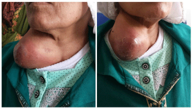
Figure 1
Figures of the mass in the supraclavicular area.
Discussion
Low-grade fibromyxoid sarcoma was described for the first time by Evans in 1987. He presented two deceptively benign-appearing, unclassifiable but very similar fibromyxoid sarcoma characterized histologically by bland, innocuous-appearing fibroblastic cells and a swirling and whorled growth pattern. He suggested the designation « low-grade fibromyxoid sarcoma » for these tumors [1]. It’s very particular morphology, its biological evolution sometimes aggressive despite a benign histological appearance, the existence of remarkable.
morphological variants, its association with specific genetic alterations and its immunophenotype make it a topic of interest to pathologists.
The Evans’ tumor occurs most often among young adults (range 20 to 50 years) but can affect people of any age, including children and elderly people. Tang reported a case of a 22-month old boy with a large LGFMS in the right cheek [3]. The male predominance appears to be a typical feature of LGFMS in most studies [2,4]. Our patient was a female aged 70 years. LGFMS occurs in the soft tissues of the extremities, but cases have been reported involving the thoracic cavity, abdomen and bowel, and peritoneum, superficial locations are rarely observed [5-8]. Localization in head and neck is very rare. Morgan reported four head and neck cases. The tumors were located in the posterior cervical spine, facial skin, mandible and larynx [4]. Tang, in a data review based on the medline database, reported only 17 head and neck cases of 245 LGFMS of deep soft tissues cases [3]. The size may vary from 1 cm to over 20 cm in diameter [5]. The case we present were located in the right supra clavicular area.
CT scan demonstrates a well-delineated, relatively low density to isodense tumor. Enhanced CT will illustrate heterogeneous enhancement; however, the enhancement is not obvious in some cases [3]. In our case, the tumor was isodense, repressing the sternocleidomastoid muscle and the right thyroid lobe, and taking the vascular carotid jugular package. The enhancement was heterogene. The magnetic resonance imaging (MRI) is admitted to be more efficient.
Histologically, LGFMS are characterized by alternating fibrous and myxoid areas, with a swirling, whorled growth pattern, at least in some parts, low to moderate cellularity, and bland, deceptively benign-appearing fibroblastic spindle cells, with no or slight nuclear pleomorphism and few mitotic figures. Enrolled vessels with hypercellularity and perivascular sclerosis are often visible.
It is more common for recurrent and metastatic neoplasms to closely resemble the primary tumor [3-5]. Sometimes, Evans’ tumor may also have collagen rosettes made of a rounded sclerotic center surrounded by a palisade of plump fibroblasts. When the tumor is particularly rich in rosettes, the designation “hyalinizing spindle cell tumor with giant rosettes” (HSCT) may be used. It is just a special morphological variant of Evans’ tumor [9].
Sometimes a "epithelioid sclerosing fibrosarcoma" is developed in an Evans’ tumor background, and this is quite obviously a different morphological variant of the disease [10]. In our case, histological exam showed a fibroadipose tissue, dissociated by a malignant mesenchymal tumor proliferation of spindle and globular cells with moderate eosinophilic cytoplasm. Nuclei were hyperchrome and irregular with many atypical figures and mitosis which is consistent with literature. In immunohistochemistry exam, LGFMS may be occasionally positive for desmin, smooth muscle actin, epithelial membrane antigen and CD34, but it is generally negative for S100 protein, Leu-7, and neuron-specific enolase. Recently, MUC4 is observed in the majority of Evans’ tumors, including HSCT and sclerosing epithelioid fibrosarcoma [11,12]. The sensitivity, but is not 100% [13]. Specificity is excellent. Immunohistochemestry exam, in our patient, was positive for cytokeratin, MUC4 and CD34. It was negative for smooth muscle lactin, desmine and PS100.
Molecularly, Evans' tumor is characterized by a translocation of chromosomes 7 and 16 or a ring chromosome juxtaposing portions of genes [14]. Wide surgical resection was considered the best treatment of LGFMS by many investigators. In addition, adjuvant radiotherapy and chemotherapy were mentioned in some reports. No evidence was found that radiotherapy or chemotherapy might cure the disease effectively. Because of the relative high rate of recurrence and distant metastasis, long time follow-up is advised [3]. Many recent larger series of LGFMS did not identify any cases in the head and neck region, which means that our case is exceptional.
Figure 2
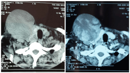
Figure 2
Axial section of the neck before and after injection of contrast product with enhancement showing the invasion of the vascular carotid jugular package, the right thyroid lobe (nodule), with an intact larynx.
Figure 3
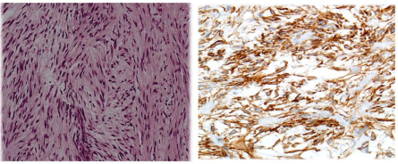
Figure 3
Representative low-grade fibromyxoid sarcoma pictures, strongly positive for MUC4 (MUC4 immunohistochemistry, X400).
Conclusion
Low grade fibromyxoid sarcoma is a rare tumor of deep soft tissues, rarely developed in head and neck. Its diagnosis is not always easy. The progress in immunohistochemistry and molecular biology may help to diagnose this deceptively bland malignancy tumor.
References
- Evans HL. Low-grade fibromyxoid sarcoma. A report of two metastasizing neoplasms having a deceptively benign appearance. Am J Clin Pathol. 1987; 88: 615-619.
- Périgny M, Dion N, Couture C, Lagacé R. [Low grade fibromyxoid sarcoma: a clinico-pathologic analysis of 7 cases]. Ann Pathol. 2006; 26: 419-425.
- Tang Z, Zhou ZH, Lv CT, Qin LY, Wang Y, Tian G, et al. Low-grade fibromyxoid sarcoma: clinical study and case report. J Oral Maxillofac Surg. 2010; 68: 873-884.
- Cowan ML, Thompson LD, Leon ME, Bishop JA. Low-Grade Fibromyxoid Sarcoma of the Head and Neck: A Clinicopathologic Series and Review of the Literature. Head Neck Pathol. 2016; 10: 161-166.
- Labonté S. Les tumeurs myxoïdes des tissus mous profonds. Annales de pathologie. 2014.
- Lee AF, Yip S, Smith AC, Hayes MM, Nielsen TO, O'Connell JX. Low-grade fibromyxoid sarcoma of the perineum with heterotopic ossification: case report and review of the literature. Hum Pathol. 2011; 42: 1804-1809.
- Park IJ, Kim HC, Yu CS, Kim JS, Jang SJ, Kim JC. Low-grade fibromyxoid sarcoma of the colon. Dig Liver Dis. 2007; 39: 274-277.
- Jakowski JD, Wakely PE. Primary intrathoracic low-grade fibromyxoid sarcoma. Hum Pathol. 2008; 39: 623-628.
- Lane KL, Shannon RJ, Weiss SW. Hyalinizing spindle cell tumor with giant rosettes: a distinctive tumor closely resembling low-grade fibromyxoid sarcoma. Am J Surg Pathol. 1997; 21: 1481-1488.
- Evans HL. Low-grade fibromyxoid sarcoma: a clinicopathologic study of 33 cases with long-term follow-up. Am J Surg Pathol. 2011; 35: 1450-1462.
- Doyle LA, Möller E, Dal Cin P, Fletcher CD, Mertens F, Hornick JL. MUC4 is a highly sensitive and specific marker for low-grade fibromyxoid sarcoma. Am J Surg Pathol. 2011; 35: 733-741.
- Doyle LA, Wang WL, Dal Cin P, Lopez-Terrada D, Mertens F, Lazar AJ, et al. MUC4 is a sensitive and extremely useful marker for sclerosing epithelioid fibrosarcoma: association with FUS gene rearrangement. Am J Surg Pathol. 2012; 36: 1444-1451.
- Linos K, Bridge JA, Edgar MA. MUC 4-negative FUS-CREB3L2 rearranged low-grade fibromyxoid sarcoma. Histopathology. 2014; 65: 722-724.
- Fletcher CDM, Bridge JA, Hogendoorn PCW, Mertens F. WHO classification of tumours of soft tissue and bone. 4th edn. Lyon: IARC; 2013.