Case Report
Carotid Body Tumor Misdiagnosed as Adenocarcinoma Metastases
Ozbay I, Oghan F*, Kucur C, Aksoy S and Erdogan O
Department of Otolaryngology, Dumlupinar University, Turkey
*Corresponding author: Fatih Oghan, Department of Otolaryngology, Medicine Faculty of Dumlupinar University, Kutahya, Turkey
Published: 07 Jul, 2016
Cite this article as: Ozbay I, Oghan F, Kucur C, Aksoy
S, Erdogan O. Carotid Body Tumor
Misdiagnosed as Adenocarcinoma
Metastases. Ann Clin Case Rep. 2016;
1: 1046.
Abstract
Forty three year old female patient was forwarded to us with the diagnosis of metastatic adenocarcinoma of unknown by the oncologist with a mass in the neck. In history, he said growing left neck mass in 5 months. Neck ultrasound (USG), neck CT and chest CT were done before coming to our clinics. Fine needle aspiration (FNA) results from mass were reported as metastatic adenocarcinoma. Patients PET was performed and hypermetabolic areas were found in the left cervical region and rectum. Two tumor biopsies were taken from rectum at different times for diagnostic and therapeutic purposes and reported as negative, therefore, neck dissection was planned. Round encapsulated mass with 3 x 2.7 x 2.5 cm was detected in left carotid bifurcation, intraoperatively. The mass was excised by working in subadventisyel. As pathology results, the mass was reported as paraganglioma, on the other hand, the neck dissection material was reported as reactive hyperplasia. Preoperative fine needle aspiration biopsy results can lead to incorrect diagnoses. The carotid body tumors can be misdiagnosed as malignancies in histopathological evaluation. We believe that the preoperative evaluation of the patient by a multidisciplinary team composed of oncologist, otolaryngologist, pathologist and radiologist will reduce this possibility.
Keywords: Carotid body tumor; Adenocarsinoma; Biopsy
Introduction
Neck swelling is quite frequent seen in various medical disciplines such as oncology, pathology,
ear-nose-throat, general surgery, internal medicine, pediatrics, thoracic surgery, radiotherapy and
creates a common interest. The neck is the busiest parts of the body's lymphatic network and very
different and a lot of neoplasm can be seen in the neck. For this reason, the differential diagnosis
is important in neck masses. In differential diagnosis; history, ultrasonography (USG), computed
tomography (CT), magnetic resonance (MR), fine needle aspiration (FNA) is utilized. FNA has
become the standard in the diagnosis and treatment of neck masses. Different opinions regarding
the success of FNA in the diagnosis has been reported in the literature [1,2]. The experience is
important for receipt, preparation and evaluation of aspiration. The most important reason for
misdiagnosis was shown as aspiration from inappropriate places and improper preparation [1,2].
FNA is a good method in the diagnosis of head and neck malignancies, especially a highly sensitive
method for the detection of metastatic carcinoma of the lymph nodes [3]. The carotid body tumors
are rare tumors developing from paraganglionic cells of the carotid body and they are often found in
common carotid artery bifurcation. The carotid body tumors are rare, asymptomatic, slow-growing
and usually benign tumors. Malignant transformation of tumor is between 3-10 % and metastasis
has been published as 2% [4,5]. Metastatic spread often happens to regional lymph nodes.
In this article, we present a patient with carotid body tumor misdiagnosed as adenocarcinoma
metastases in the light of the literature.
Case Presentation
Forty-three-year-old female patient with a mass in the neck was forwarded to us by oncologist
with the diagnosis of metastatic adenocarcinoma of unknown. In history, he said growing left neck
mass in 5 months. Neck ultrasound (USG), neck CT and chest CT were done before coming to
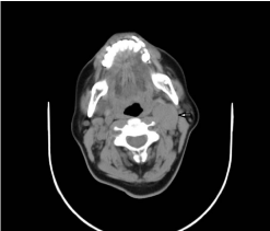
our clinics. In the neck CT, there was a ovoid, smooth contours of approximately 30x20x45 mm
in size and isodense mass pushing the left internal carotid artery (ICA) posteriorly, and locating in
the posterior of the sternocleidomastoid (SCM) muscle (Figure 1). Fine needle aspiration (FNA)
results from mass were reported as metastatic adenocarcinoma. Patients PET was performed and
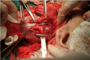
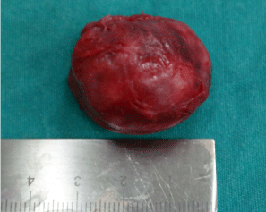
hypermetabolic areas were found in the left cervical region and rectum. Two tumor biopsies were taken from rectum at different times for diagnostic and therapeutic purposes and reported as negative, therefore, neck dissection was planned. Round encapsulated mass with 3 x 2.7 x 2.5 cm was detected in left carotid bifurcation, intraoperatively. The mass was excised by working in subadventisyel (Figure 2 and 3). As pathology results, the mass was reported as paraganglioma, on the other hand, the neck dissection material was reported as reactive hyperplasia.
Postoperative complications were not observed in the patient. Although flushing episodes were observed in the patient's in first postoperative days, these complaints were disappeared in follow-up.
Figure 1
Figure 2
Figure 3
Discussion
Neck mass is a common pathological condition involving many medical disciplines. All tumors should be approached with a suspected neck malignancy until proved otherwise, and primary studies should be performed in order to determine the pathology taking into account the possibility of a metastasis. Therefore, detailed ENT examination in the approach of the neck masses is vital especially in metastatic neoplasms. It is not possible to identify the primary focus without a complete ear, nose, and throat examination. Multidisciplinary approach should be followed in the diagnosis of mass such as radiology, pathology, oncology branches. In our case, oncology directed the patient to our clinics in the diagnosis stage; FNA diagnosed incorrectly, preliminary results of PET / CT results have led us to have the wrong directions.
The staging of head and neck tumors is an important part of diagnosis and treatment. Especially, in locally advanced head and neck cancer are about 10% in the distant metastatic disease [6]. PET / CT provide both local staging and showing distant metastases and it is a good opportunity in this regard affecting the patient management. In a study, especially, high-risk patients were selected and evaluated for synchronous and metachronous metastases of patients as the first staging of head and neck cancer and it was conducted in F-18 FDG PET / CT. The sensitivity and specificity for distant metastasis were 96.8% and 95.4%, respectively, [7]. In our case PET / CT results, hyper metabolic areas of the rectum were followed and rectal adenocarcinoma is thought to be inducing metastasis in the neck. Tumor biopsies were taken from the rectum twice and have resulted as negatives in both of them. Although PET / CT have high reliability, fallibility should be kept in mind.
Head and neck masses develop due to inflammatory, infectious, benign or malignant neoplasms, and cystic lesions. The story of the patient and clinical findings are necessary for diagnosis [8]. However, morphological examination is necessary in order to make a definitive diagnosis in most cases. In addition, during preoperative evaluation, accurate diagnosis is of paramount importance in determining the surgical and technical technique [9]. Therefore, importance of FNA in the diagnosis and treatment planning increases. FNA is cheap, reliable and easy diagnostic methods to apply. However, different opinions in the literature regarding the diagnosis success of FNA has been reported [1,2]. In a study reported by Ozbay et al. [10] regarding to non thyroidal FNA results containing all head and neck masses, they reported 68 patients with 95.1% accuracy, 97.5% sensitivity, 92.8% specificity, 1.5% false positive and 2.9% false negative results. In our case, treatment plans has changed completely due to wrong result as adenocarcinoma metastasis.
In the histopathological basis of carotid body tumor, the majority of main cells and supporting cells that surround them create "zellball island". Main cells contain cell nuclei with fine chromatin network structure and amfofilik or eosinophilic cytoplasm. These cells are sometimes as short cords in carcinoid tumors, sometimes gland-like structures as in adenocarcinoma, and sometimes rosette-like structures as seen in neuroblastic tumors [11]. Therefore, histological differential diagnosis is required in cytological differential diagnosis of these tumors. Our patient was misdiagnosed as metastasis of adenocarcinoma. This kind of differential diagnosis to avoid misdiagnosis is necessary to rule out the possibilities.
The carotid body tumor is caused by chemoreceptor tissue in the carotid bifurcation. It is frequently benign and non functional. Growing mass in the anterior neck triangle should raise this pathology. Due to the slow development, it is often asymptomatic until it reaches a certain size [12]. They remain silent until they press cranial nerve or vascular structures. In patients with functional, it can occur with symptoms related to the release of catecholamines. It is more common in women and at high altitudes. They are tend to be seen together with other cervical paraganglioma of carotid body tumors, head and neck and other malignant paraganglioma (lung, larynx, breast carcinoma) and are tend to be bilateral or multiple. Therefore, each patient with unilateral mass should be examined carefully including all neck and thorax [13,14]. In our case, it wasasymptomatic left neck mass growing in five months, and it has no symptoms, no family history. There was no other focus showed by the body scan.
The choice of therapy for carotid body tumor should be planned considering patient's symptoms, age, tumor size, growth rate and complications of the surgery. Radiotherapy and / or embolization can be selected because it has slow growth pattern, low malignancy and surgical treatment risk [15]. In our case, the mass was excised by working in subadventisyel and postoperative complications were not observed in the patient.
Conclusion
Unknown primary tumors of the neck should be evaluated multidisciplinary. A detailed ENT examination and all attempts must be carried out carefully in order to find primary tumor. FNA is one of the diagnostic phase, therefore it should be done professionally and should be reported by a good cytologist. Otherwise, patients with suspected cancer may be uneasy for months like our patient. The patient waited 3-4 months until reaching surgical pathology report as benign. If the patient was admitted to our clinic directly and FNA results was diagnosed correctly, the patient would not be uneasiness for 3-4 months.
References
- Lee KR, Foster RS, Papillo JL. Fine needle aspiration of the breast. Importance of the aspirator. Acta Cytol. 1987; 31: 281-284.
- Shah SB, Singer MI, Liberman E, Ljung BM. Transmucosal fine-needle aspiration diagnosis of intraoral and intrapharyngeal lesions. Laryngoscope. 1999; 109: 1232-1237.
- el Hag IA, Chiedozi LC, al Reyees FA, Kollur SM. Fine needle aspiration cytology of head and neck masses. Seven years' experience in a secondary care hospital. Acta Cytol. 2003; 47: 387-392.
- Hallett JW, Nora JD, Hollier LH, Cherry KJ, Pairolero PC. Trends in neurovascular complications of surgical management for carotid body and cervical paragangliomas: a fifty-year experience with 153 tumors. J Vasc Surg. 1988; 7: 284-291.
- Nora JD, Hallett JW, O'Brien PC, Naessens JM, Cherry KJ, Pairolero PC. Surgical resection of carotid body tumors: long-term survival, recurrence, and metastasis. Mayo Clin Proc. 1988; 63: 348-352.
- Black RJ, Gluckman JL, Shumrick DA. Screening for distant metastases in head and neck cancer patients. Aust N Z J Surg. 1984; 54: 527-530.
- Haerle SK, Schmid DT, Ahmad N, Hany TF, Stoeckli SJ. The value of (18)F-FDG PET/CT for the detection of distant metastases in high-risk patients with head and neck squamous cell carcinoma. Oral Oncol. 2011; 47: 653-659.
- Schelkun PM, Grundy WG. Fine-needle aspiration biopsy of head and neck lesions. J Oral Maxillofac Surg. 1991; 49: 262-267.
- Önder T, Aktas D, Günhan Ö, Özkaptan Y. Bas ve Boyun Kitlelerinde Ince Igne Aspirasyon Biyopsisi. K.B.B. ve BBC Dergisi. 1994; 2: 32-37.
- Özbay AS, Çiftçioglu MA, Sütbeyaz Y, Aktan B, Ertas A. Boyun kitlelerinde ince igne aspirasyon biopsisi sonuçlarimiz. KBB ve Bas Boyun Cerrahisi Dergisi 1994; 2: 226-230.
- Weiss SW, Goldblum JR. Enzinger and Weiss’s Soft Tissue Tumors, 4 th ed. St.Louis: Mosby. 2001; 1323-1360.
- Rodríguez-Cuevas S, López-Garza J, Labastida-Almendaro S. Carotid body tumors in inhabitants of altitudes higher than 2000 meters above sea level. Head Neck. 1998; 20: 374-378.
- Ünlü Y, Azman A, Özyazicioglu A, Becit N, Erol K, Ceviz M, et al. Carotid body tumours (Paragangliomas). Asian Cardivasc Thorac Surg. 2001; 3: 208-211.
- Elmaci TT, Kargi A, Onursal E. Karotis paragangliomalari. Damar Cer Derg. 1999; 3: 111-115.
- Erentug V, Bozbuga NU, Sareyyüpoglu B ve ark. Karotis Cisim Tümötlerinde Cerrahi Yaklasimlar. Türk Gögüs Kalp Damar Cerrahisi Dergisi. 2004; 12: 277-279.


