Case Report
Alveolar Rhabdomyosarcomatous Endometrial Polyp in a 70 Year Old Woman
Niranjanakesav1, Sathiya Bama2*, Radhakrishnanm,1 and Arbind Kumar3
1Department of Pathology, Virchow Biopsy Centre, India
2Department of Pathology, IRT Perundurai Medical College and Hospitals (IRT-MPCH), India
3Department of Pharmacology, IRT Perundurai Medical College and Hospitals (IRT-MPCH), India
*Corresponding author: Sathiya Bama, Department of Pathology, IRT Perundurai Medical College and Hospitals (IRT-MPCH), Perundurai, 638053, Tamil Nadu, India
Published: 28 Jun, 2018
Cite this article as: Niranjanakesav, Sathiya Bama,
Radhakrishnan, Kumar A. Alveolar
Rhabdomyosarcomatous Endometrial
Polyp in a 70 Year Old Woman. Ann
Clin Case Rep. 2018; 3: 1526.
Abstract
This case report concerns a 70 year woman with a complaint of abnormal vaginal bleeding and foul smelling vaginal discharge of a short duration. Speculum examination revealed a huge polyp occupying the cervix and extending further downwards. On operation, it turned out to be an endometrial polyp arising from the fundus. Histologically the polyp showed classical features of alveolar rhabdomyosarcoma; confirmed immunochemically.
Introduction
Rhabdomyosarcoma of uterus is an unusual sarcomatous tumor in gynecologic practice; and when seen it is associated as a component of carcinosarcoma; considered now as a classical example of epithelial mesenchymal transition tumor in human.
Oncogenesis
As a distinct tumor, this is recognized long back in 1968 by Enter line and Horn [1]. Later was analyzed in detail a large survey of 112 cases by Enzinger and Shiraki [2]. The oldest patient in their series was 58 years. Parham and Barr [3] succinctly defined it as cellular sarcomatous neoplasm; containing a monomorphous population of primitive cells and showing skeletal muscle differentiation.
Case Presentation
The main complaints of this 70 year old lady was foul smelling vaginal discharge for few months and post menopausal bleeding for a month. Speculum revealed a fungating mass protruding into the cervix and vagina. Subsequently a total abdominal hysterectomy with BSO was done. A large polypoidal mass (5 cm×5 cm×4 cm); fleshy in nature was arising from fundus and occupying the entire uterus and cervix. There were two other smaller polyps, were friable, necrotic and partly gangrenous at its tip. Microscopically it was moderately cellular; separated by thick fibrovascular septa. Pseudo alveolar spaces were seen throughout this neoplasm, constituting more than 50% of the tumor showing features consistent with alveolar rhabdomyosarcoma (ARMS) (Figure 1). The non cohesive cells contained in these spaces are mostly nondescript. Classical rhabdomyoblasts with round morphology and abundant eosinophilic cytoplasm were noticed in their midst, strap and racquet shaped cells being rare. Bulbous cells were arranged at the periphery sticking to the walls of septa. In addition, in few sections a spindle shaped sarcomatous growth; with fasciculated pattern co-existed with the above but not mixing with predominantly primitive cells featuring leiomyosarcoma (LMS). This collison tumor showed multiple foci of coagulative necrosis with a mitotic rate of 10 per 10 high power field. The abrupt transition between ARMS and low grade LMS was surprisingly distinct and morphologically heterogenous (Figure 2). Immunochemistry was done to confirm the nature of the neoplastic cells; proving thereby the histogenesis of this tumor conclusively. The tumor cells were strongly positive for skeletal muscle markers like cytoplasmic desmin, muscle specific actin and nuclear positivity for myogenin D. Cytokeratin was positive for benign endometrial glands; distributed singly amidst malignant cells (Figures 3).
Discussion
Endometrial polyps at this age are unusual; most often are due to Malignant Mixed Mullerian
Tumor (MMMT) or rarely due to adenocarcinoma arising from EIN in endometrial polyp. The
rarity of this tumor is well illustrated in the IRS study dealing with ARMS; in which only eight such cases were of genitourinary system [4]. The histological hallmark of this neoplasm of pseudo-alveolar spaces amidst solidly
arranged tumor cells; after downgrading its extent from 75% to 50%
helped to diagnose this condition better and realistically. Another
histological feature like detecting many forms of rhabdomyoblasts
amidst nondescript cells arranged solidly as well as in pseudo alveolar
spaces helped to diagnose this condition accurately and rule out other
possibilities like small cell synovial sarcoma, malignant lymphoma,
small blue cell tumors and solid carcinomas of epithelial origin.
After the advent of immunochemistry, the diagnosis can be firmly
established by employing myogenic transcription factors; myogenin
and Myo-D1. They are transcriptional regulatory proteins expressed
early in muscle differentiation [5] and display a nuclear staining
pattern. In a recent review of 956 cases, myogenin and MyoD1 showed
a sensitivity of 97% and specificity of 90% and 95% respectively [6].
Myogenin is considered technically superior to other markers [7].
Though Ober long ago recognized malignant mesenchymal tumors of
uterus as homologous or hetero logous or mixed, the number of case
reported as mixed is still rare; particularly collison tumor as described
in this patient.
Reported cases of ARMS involving uterus are rare and when
compared with the case under discussion differed markedly in
presentation. Fukanya [8] reported a case of ARMS of uterine corpus
in a 70 year old woman with the tumor replacing the wall of the
body of uterus by multiple friable nodules. She developed extensive
metastases and died 12 months after surgery. The second such case
[9] was in a 21yr old lady; unique for uterine inversion secondary to
ARMS. The last such case was seen in a 44 year old African woman
[10]; presenting with widespread disseminated tumor and involving
all abdominal viscera. It is clear the case details recorded in our patient
differed vastly from other reported cases and appears unique. We feel
from our experience myogenin immunochemistry in future will help
to discover many more cases and to plan for effective therapy in such
patients.
Figure 1
Figure 1
a) Low power view of ARM b) high power view to show
rhabdomyoblastic differentiation.
Figure 2
Figure 2
Medium power view of leiomyosarcoma.
Figure 3
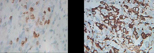
Figure 3
a) Myogenin nuclear positivity b) Desmin cytoplasmic positivity
square.
References
- Lopez Garcia, Palacios J. Pathologic and molecular features of uterine carcinosarcomas. Seminars in Diag Path 2010; 27(4): 274-86.
- Enzinger FM, Shiraki. Alveolar Rhabdomyosarcoma. An analysis of 110 cases. Cancer.1969; 24(1):18-31.
- Parham DM, Barr FG. Alveolar Rhabdomyosarcoma. Christopher DM.Fletcher, Julia Bridge, Pancras CW, Hogendron and Fredrik Mertens. WHO classification of tumours of soft tissue and Bone. Lyon: IARC Press;2002; 130-132.
- Goldblum Tr, Folpe AC, Weiss SW. Soft tissue tumors, Elsevier, 6th Ed; 2013;604.
- Hornick JL. Practical soft tissue Pathology. A diagnostic approach. Hornick; Elsevier; 2013; 207.
- Hosit H, Sugimolo T, Hayashi y. Differential expression of myogenic regulatory proteins. MyoD1 and myogenin in human RMS. Int.T. Cancer 1992; 50: 977-983.
- Shunjin Cui, Hiroshi Hano, Tohru Harada, Shigeharu Takai, Fumiaki Masui, Shinichiro Ushigome, et al. Evulation of new monoclonal Myo-D1 and anti-myogenin antibodies for the diagnosis of Rhabdomyosarcoma. Pathol. Int.1999; 49(1): 62-8.
- Fukunaga,M. Pure alveolar rhabdomyosarcoma of uterine corpus. Pathol Int. 2011; 61(6): 377-81.
- Case AS, Kirby TO, Conner MG, Huh WK, et al. A case report of rhabdomyosarcoma of uterus associated with uterine inversion. Gynecol.Oncol. 2005;96(3): 850-3.
- A T Odoi, E T Dassah, D E Darkey, O Owusu-Afriyie, A Y Valkov. Advanced Alveolar Rhabdomyosarcoma of the Uterus: A Case Report African Journal of Reproductive Health. 2009;13(1): 167-73.