Case Report
Abdominal Splenosis Mimicking Peritoneal Spread of Prostate Adenocarcinoma in 68Ga- PSMA PET/CT- A Case Report
Ron Y*, Kesler M, Lerman H and Even-Sapir E
Department of Nuclear Medicine, Tel-Aviv Sourasky Medical Center, Israel
*Corresponding author: Yosef Ron, Department of Nuclear Medicine, Tel Aviv Sourasky Medical Center, 6 Weizmann St. Tel Aviv, Israel
Published: 17 Jun, 2018
Cite this article as: Ron Y, Kesler M, Lerman H, Even-Sapir
E. Abdominal Splenosis Mimicking
Peritoneal Spread of Prostate
Adenocarcinoma in 68Ga- PSMA PET/
CT- A Case Report. Ann Clin Case Rep.
2018; 3: 1520.
Abstract
Background: 68Ga-labeled prostate-specific membrane antigen ligand (68Ga-PSMA) PET/CT has
become a routine in our department for imaging patients with prostate cancer, staging of medium
and high-risk patients, assessment of biochemical failure and planning of 177Lu-PSMA therapy. Data
accumulated exposes us to potential pitfalls in interpretation of 68Ga-PSMA PET/CT studies. We
report a case of a patient with potential misinterpretation of peritoneal spread.
Case Presentation: A 69-years-old man patient with prostate adenocarcinoma Gleason 9 underwent
68Ga-PSMA PET/CT for disease extent assessment. Multiple soft tissue masses accumulating the
tracer were detected in the left subdiaphragmatic area, anterior abdomen adjacent to the abdominal
wall and on the bowel serosa presenting as abdominal metastatic spread. However, the spleen was
not identified in its usual anatomical place. We approached the patient revealing that he had a
post-traumatic splenectomy two decades earlier. 99mTc-labeled heat-damaged red blood cells (99mTcdRBCs)
SPECT/CT study confirmed our interpretation of 68Ga-PSMA PET/CT as identifying
physiological tracer uptake in abdominal splenosis rather than metastatic spread.
Conclusion: Splenosis and not metastasis as the cause for abdominal lesions showing increased
68Ga-PSMA uptake, should be considered in a patient with a previous history of abdominal trauma
or splenectomy where spleen is not identified in its normal bed. Correlation with 99mTc-dRBCs scan
may assist in localizing ectopic splenic tissue sites.
Keywords: PET; Prostate adenocarcinoma; 68Ga-PSMA; Splenosis
Introduction
Prostate cancer is the second most frequently diagnosed cancer in men and the fifth leading cause of cancer death worldwide [1]. PET imaging with 68Ga-PSMA ligands has been recently introduced in the imaging algorithm of patients with prostate cancer [2]. The spleen is a PSMA-expressing organ showing physiologic uptake of the tracer [3,4,5]. We report a case of post splenecomy abdominal splenosis as a potential cause for pitfall on 68Ga-PSMA PET/CT by abdominal metastatic spread. 99mTc-dRBCs SPECT/CT study has been used for re-assuring the diagnosis of splenosis. We also review other unexpected settings of benign PSMA expressing tissue as potential pitfalls on 68Ga- PSMA PET/CT interpretation.
Case Presentation
A 69-years-old male patient with a newly-diagnosed Gleason 9(5+4) prostate adenocarcinoma,
was referred for staging with 68Ga-PSMA PET/CT. Serum level of PSA was 1.68 ng/ml. Eight months
earlier the patient underwent Transurethral Resection of the Prostate (TURP) due to Benign prostatic
hyperplasia. PET/CT was acquired following administration of 156 MBq (4.2 mCi) 68Ga-PSMA. In
addition to uptake at the primary tumor site at the prostate gland, multiple foci of increased tracer
uptake SUVmax of 4.15 ± 1.15 was detected corresponding to multiple soft tissue masses ranging in
size between 8.0 mm to 40.0 mm at the left abdomen subdiaphragmatically, at the anterior abdomen
adjacent to the abdominal wall and on the transverse colon serosa (Figure 1).
Absence of normal spleen on CT has raised the suspicion of post splenectomy splenosis. Patient's
history revision has revealed a post-traumatic splenectomy two decades earlier. A subsequent 99mTcdRBCs
scan, planar and SPECT/CT was performed using a hybrid SPECT/CT camera (Optima 640,
GE Healthcare) (Figure 2).
There was a full matching between 99mTc-dRBCs and 68Ga-PSMA
uptake in the abdominal lesions confirming that the lesions seen
on PET/CT represented physiologic uptake in splenosis and not
metastatic spread (Figures 3-5).
Figure 1

Figure 1
A whole-body 68Ga-PSMA PET/CT study Maximum Intensity Projection (MIP) (A), coronal CT, PET and fused PET/CT (B, C and D respectively) and
axial CT, PET and fused PET/CT (E, F and G respectively), demonstrated a tracer uptake (SUVmax, 4.15 ± 1.15) by multiple soft tissue masses in the left abdomen,
below the diaphragm, left mesentery and the anterior abdomen at the navel level (white and black arrows).
Figure 2
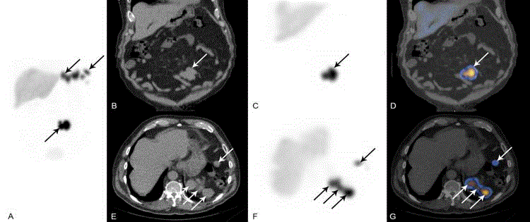
Figure 2
A subsequent 99mTc-dRBCs SPECT/CT study Maximum Intensity Projection (MIP) (A), coronal CT, SPECT and fused SPECT/CT (B, C and D respectively)
and axial CT, SPECT and fused SPECT/CT (E, F and G respectively), revealed multiple areas of increased activity, anatomically matching with the suspected
masses seen on the 68Ga-PSMA PET/CT study, confirming the diagnosis of splenosis.
Discussion
There is accumulating clinical data showing a high diagnostic accuracy of the novel 68Ga-PSMA PET/CT technique for staging of patients with newly diagnosed intermediate and high-risk prostate cancer. Diagnostic accuracy of the study interpretation is based on familiarity with potential pitfalls caused by unexpected sites of physiologic uptake or uptake with non-tumoral lesions. Several previous publications presented cases of increased 68Ga-PSMA uptake in benign lesions including adrenal adenomas, paravertebral schwannoma, Paget's bone disease, splenic sarcoidosis, follicular thyroid adenoma, meningioma, serous cystadenoma of the pancreas, Pseudoangiomatous Stromal Hyperplasia (PASH) of the breast, liver hemangioma, subacute stroke and newly formed blood vessels, tuberculosis and Healing nonmalignant fracture [2,6-16]. Celiac lymph nodes were reported to show physiologic uptake and so were PSMA-expressing organs such as spleen [2,3,4]. Therefore as shown in the current presented patient, ectopically located splenic tissue may lead a false-positive misinterpretation of PET/CT confusing ectopic sites of normal splenic tissue with physiologic tracer uptake and metastatic spread. Ectopic splenic tissue is manifested in two distinct forms, accessory spleens and splenosis. Accessory spleens are congenital [17], while splenosis may be found following trauma in 26% to 65% of cases and following elective splenectomy for hematologic disorders in 16% to 20% [18]. Splenosis detection is usually incidental, and no therapy is indicated when asymptomatic. It can be found with any shape and size, usually with no expression of typical architecture such as hilum or defined capsule [19]. Splenosis is of clinical significance in the case of bleeding, local compression on adjacent structures and when misdiagnosed as tumor [20-22]. 99mTcdRBCs imaging is a highly specific since the tracer is taken almost exclusively by splenic tissue [23,24]. It is a sensitive imaging technique given the high contrast between splenules and surrounding tissues. Performing the scan using a hybrid SPECT/CT camera allows both, diagnosing the tissue of the lesion in question as composed of splenic tissue and locating the ectopic splenic tissue in the abdomen [25]. In the presented case SPECT/CT and PET/CT could be correlated for each suspected lesion.
Figure 3
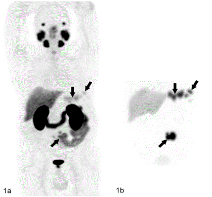
Figure 3
Maximum Intensity Projection (MIP) of 68Ga-PSMA (PET/CT) (A),
and 99mTc-dRBCs (SPECT/CT) (B). There was a full matching between 99mTcdRBCs
and 68Ga-PSMA uptake in the abdominal lesions, confirming that the
lesions seen on PET/CT represented physiologic uptake in splenosis and not
metastatic spread.
Figure 4
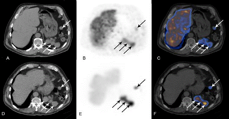
Figure 4
68Ga-PSMA PET/CT axial CT, PET and hybrid PET/CT (A, B and C respectively), and 99mTc-dRBCs SPECT/CT axial CT, SPECT and hybrid SPECT/CT
(D, E and F respectively), demonstrated the suspected masses to match anatomically.
Figure 5
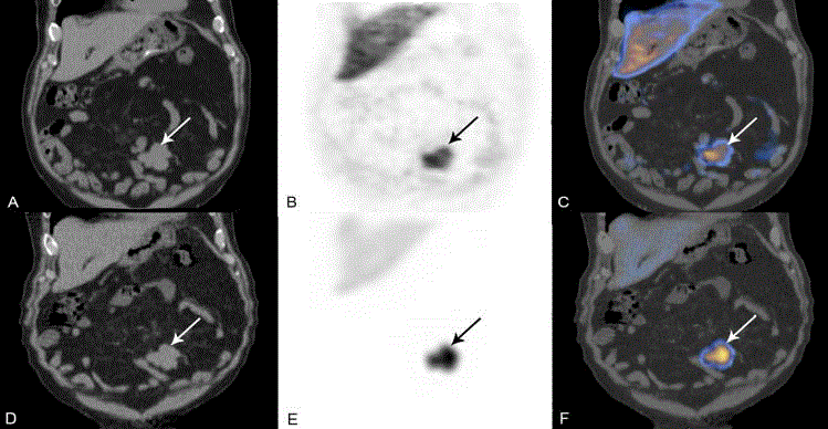
Figure 5
68Ga-PSMA PET/CT coronal CT, PET and hybrid PET/CT (A, B and C respectively), and 99mTc-dRBCs SPECT/CT coronal CT, SPECT and hybrid
SPECT/CT (D, E and F respectively), demonstrated the suspected masses to match anatomically.
Conclusion
Abdominal sites of 68Ga-PSMA uptake may be detected in case of splenosis. These lesions may be misinterpreted as metastatic abdominal spread in patients with prostate adenocarcinoma. Previous patient's history and absent spleen in the normal location should raise the possibility of splenosis. The latter can be confirmed by a complementary 99mTc-dRBCs scan.
References
- Jemal A, Bray F, Center MM, Ferlay J, Ward E, Forman D. Global cancer statistics. CA Cancer J Clin. 2011;61(2):69-90.
- Rauscher I, Maurer T, Fendler WP, Sommer WH, Schwaiger M, Eiber M. 68Ga-PSMA ligand PET/CT in patients with prostate cancer: How we review and report. Cancer Imaging. 2016;16:14.
- Weineisen M, Schottelius M, Simecek J, Baum RP, Yildiz A, Beykan S, et al. 68Ga- and 177Lu-labeled PSMA I&T: Optimization of a PSMA-targeted theranostic concept and first proof-of-concept human studies. J Nucl Med. 2015;56(8):1169-76.
- Demirci E, Sahin OE, Ocak M, Akovali B, Nematyazar J, Kabasakal L. Normal distribution pattern and physiological variants of 68Ga-PSMA-11 PET/CT imaging. Nucl Med Commun. 2016;37:1169-79.
- Prasad V, Steffen IG, Diederichs G, Makowski MR, Wust P, Brenner W. Biodistribution of [(68)Ga]PSMA-HBED-CC in patients with prostate cancer: Characterization of uptake in normal organs and tumour lesions. Mol Imaging Biol. 2016;18(3):428-36.
- Law WP, Fiumara F, Fong W, Miles KA. Gallium-68 PSMA uptake in adrenal adenoma. J Med Imaging Radiat Oncol. 2016;60(4):514-7.
- Rischpler C, Maurer T, Schwaiger M, Eiber M. Intense PSMA-expression using 68Ga-PSMA PET/CT in a paravertebral schwannoma mimicking prostate cancer metastasis. Eur J Nucl Med Mol Imaging. 2016;43(1):193-4.
- Artigas C, Alexiou J, Garcia C, Wimana Z, Otte FX, Gil T, et al. Paget bone disease demonstrated on 68Ga-PSMA ligand PET/CT. Eur J Nucl Med Mol Imaging. 2016;43(1):195-6.
- Kobe C, Maintz D, Fischer T, Drzezga A, Chang DH. Prostate-specific membrane antigen pet/ct in splenic sarcoidosis. Clin Nucl Med. 2015;40:897-8.
- Kanthan GL, Drummond J, Schembri GP, Izard MA, Hsiao E. Follicular thyroid adenoma showing avid uptake on 68 Ga PSMA-HBED-CC PET/CT. Clin Nucl Med. 2016;41(4):331-2.
- Bilgin R, Ergül N, Çermik TF. Incidental meningioma mimicking metastasis of prostate adenocarcinoma in 68Ga-labeled PSMA ligand PET/CT. Clin Nucl Med. 2016;41(12):956-8.
- Chan M, Schembri GP, Hsiao E. Serous cystadenoma of the pancreas showing uptake on 68Ga PSMA PET/CT. Clin Nucl Med. 2017;42(1):56-7.
- Malik D, Basher RK, Mittal BR, Jain TK, Bal A, Singh SK. 68Ga-PSMA Expression in pseudoangiomatous stromal hyperplasia of the breast. Clin Nucl Med. 2017;42(1):58-60.
- Bhardwaj H, Stephens M, Bhatt M, Thomas PA. Prostate-specific membrane antigen pet/ct findings for hepatic hemangioma. Clin Nucl Med. 2016;41(12):968-9.
- Noto B, Vrachimis A, Schäfers M, Stegger L, Rahbar K. Subacute stroke mimicking cerebral metastasis in 68Ga-PSMA-HBED-CC PET/CT. Clin Nucl Med. 2016;41(10):e449-e51.
- Gykiere P, Goethals L, Everaert H. Healing sacral fracture masquerading as metastatic bone disease on a 68Ga-PSMA PET/CT. Clin Nucl Med. 2016;41(7):e346-347.
- Halpert B, Gyorkey F. Lesions observed in accessory spleens of 311 patients. Am J Clin Pathol 1959;32(2):165-8.
- Schrier SL. Approach to the adult patient with splenomegaly and other splenic disorders.
- Lai T, Meng C. Silent pelvic splenosis: case report. Int J Surg Case Rep. 2015; 13:129-30.
- Conway AB, Cook SM, Samad A, Attam R, Pambuccian SE. Large platelet aggregates in endoscopic ultrasound-guided fine-needle aspiration of the pancreas and peripancreatic region: A clue for the diagnosis of intrapancreatic or accessory spleen. Diagn Cytopathol. 2013;41(8):661-72.
- Yu H, Xia L, Li T, Ju M, Liu L, Wu Z, et al. Intrahepatic splenosis mimicking hepatoma. BMJ Case Rep 2009;2009:bcr06.2008.0230.
- Younan G, Wills E, Hafner G. Splenosis: A Rare Etiology for Bowel Obstruction-A Case Report and Review of the Literature. Case Rep Surg. 2015; 2015: 890602.
- Hagman TF, Winer-Muram HT, Meyer CA, Jennings SG. Intrathoracic splenosis: superiority of technetium Tc99m heat-damaged RBC imaging. Chest. 2001;120(6):2097-8.
- Henkin RE, et al. Nuclear Medicine: 2-Volume Set. Mosby; 2nd ed: Sylvester JM. 2006;954-6.
- Atkins HL, Goldman AG, Fairchild RG, Oster ZH, Som P, Richards P, et al. Splenic sequestration of 99mTc labeled, heat treated red blood cells. Radiology. 1980;136(2):501-3.