Case Report
Hemorrhagic Abdominal Mass Presenting as Shoulder Pain in the Emergency Department
Jeanine Awad Spirtos*
Department of Emergency, Cleveland Clinic Akron General, USA
*Corresponding author: Jeanine Awad Spirtos, Department of Emergency, Cleveland Clinic Akron General, 1 Akron General Avenue, Akron, Ohio, 44307, USA
Published: 03 Mar, 2018
Cite this article as: Spirtos JA. Hemorrhagic Abdominal
Mass Presenting as Shoulder Pain in
the Emergency Department. Ann Clin
Case Rep. 2018; 3: 1505.
Abstract
Shoulder pain is a common complaint in the Emergency Department (ED) and it is often secondary
to musculoskeletal causes. Patients who have multiple presentations to the ED with a similar chief
complaint and no indications of musculoskeletal etiology should be evaluated for differential
diagnosis based on the potential for referred pain. We present a case of a patient who had several
ED and outpatient visits over the course of 18 months for shoulder pain. Initial imaging studies were
unremarkable and the patient was discharged with instructions for conservative pain management.
The patient developed abdominal pain several months after the onset of shoulder pain, prompting
further investigation. Abdominal and pelvic CT scans revealed a large intra-abdominal tumor
herniating the patient’s diaphragm. The shoulder pain was determined to be secondary to
diaphragmatic irritation. The patient underwent emergent surgery to remove the mass from the
adjacent structures. The final pathology for tumor was positive for synovial sarcoma.
Keywords: Referred pain; Synovial cell carcinoma; Shoulder Pain
Case Presentation
Shoulder pain is a very common complaint in the Emergency Department (ED). The anatomy
of the shoulder permits extensive movement [1]. This potential for movement generates instability
in the joint, leading to increased risk for injury [2]. When patients have a benign physical exam and
negative imaging for musculoskeletal etiologies in the ED, they are usually discharged home with
instructions for pain control and outpatient follow up. If a patient presents on several visits with
worsening shoulder pain that is unresponsive to conservative therapy, medical providers should
consider broadening their differential beyond musculoskeletal causes of pain.
A 50 year-old female presented to the ED with shoulder pain. The patient would proceed to have
several ED and outpatient visits over an 18-month span with the complaint of left shoulder pain.
Additionally, the patient was admitted for anemia and required a blood transfusion four months
prior. At that time, there were no obvious sources of bleeding. The patient was discharged home
with a diagnosis of aplastic anemia.
During her most recent ED visit, the patient reported worsening left shoulder pain. She had
recently undergone an outpatient MRI of her left shoulder, which reportedly showed arthritis.
Results from the MRI were not available for review. She was undergoing conservative treatment for
bursitis and arthritis at that time. The patient stated that her shoulder pain had been worsening and
it was unresponsive to treatment with anti-inflammatories and narcotic pain medication.
The patient also reported a four-month history of worsening epigastric abdominal pain. The
patient stated that she had recently undergone a colonoscopy and esophagogastroduodenoscopy.
According to the patient, the results of these procedures were within normal limits. There were
no records available from either of these procedures and there was no prior abdominal imaging at
our institution. The remainder of the patient’s past medical history and past surgical history was
unremarkable.
The physical exam revealed diffuse abdominal tenderness, worst in the epigastrium. Examination
of the patient’s left shoulder was unremarkable with no overt signs of musculoskeletal pathology or
reproducible pain with palpation or range of motion. Laboratory evaluation showed a decrease in
the patient’s hemoglobin by nearly 3 gm/dL when compared to the hemoglobin drawn ten days
prior. CT scan of abdomen and pelvis showed a large heterogeneous mass measuring greater than
19 cms in the left upper abdomen herniating into the left hemidiaphragm and left thoracic cavity
with suspected active hemorrhage within the mass (Figure 1 and 2). The patient was admitted to general surgery and she underwent emergent extensive abdominal and thoracic surgery to remove the mass from the adjacent structures.
Final pathology was positive for synovial sarcoma arising from the
left abdomen.
Synovial sarcoma is a rare soft-tissue sarcoma that originates
from primitive mesenchymal cells. Tumors normally present in the
lower extremities, commonly in the popliteal fossa; however, there
are several documented cases of synovial sarcoma arising in the chest wall, heart, mediastinum, lungs, head, neck, and other unusual
sites. Intra-abdominal synovial sarcomas are rare and patients often
experience symptoms for years before diagnosis [3].
Patients can have referred shoulder pain arising from multiple
etiologies. Extrinsic causes of shoulder pain include diaphragmatic
irritation, apical lung tumors, pneumonia, myocardial ischemia,
cervical nerve root compression or metastases [4]. When evaluating
for extrinsic causes of shoulder pain, the provider must obtain a
detailed history and physical examination. Important clues pointing
to a cause other than a primary musculoskeletal disease include a
history of cancer, weight loss, rash, neck pain, chest pain, respiratory
symptoms and neurologic symptoms [5]. If the patient’s shoulder
pain is secondary to referred pain, it is often difficult to localize and
reproduce. It is also often unaffected by palpation or range of motion
[4].
Figure 1
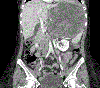
Figure 1
This is a coronal image of the CT abdomen and pelvis. The red
arrow shows the intraabdominal mass herniating through the diaphragm into
the left thoracic cavity with suspected active hemorrhage.
Figure 2
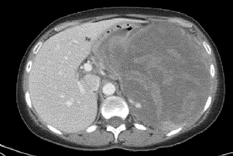
Figure 2
This is an axial image of the CT abdomen and pelvis. The red
arrow shows the heterogeneous, predominantly cystic mass in the left upper
abdomen measuring greater than 19 cms.
Conclusion
It is important that emergency medicine physicians evaluate for alternative etiologies of pain, taking into special consideration the possibility of referred pain. Evaluating for potential causes of referred pain is especially important in cases of repeated emergency department visits for the same complaint unresponsive to traditional therapies.
References
- Ramponi D. Shoulder pain. Adv Emerg Nurs J. 2011;33:114-126.
- Woodward T, Best T. The painful shoulder: Part 1. Clinical evaluation. Am Fam Physician. 2000;61: 3079-3088.
- Bakri A, Shinagare A, Krajewski K. Howard S, Jagannathan J, Hornick J, et al. Synovial sarcoma: Imaging features of common and uncommon primary sites, metastatic patterns and treatment response. AJR Am J Roentgenol. 2012;199:W208-W215.
- House J, Mooradian A. Evaluation and management of shoulder pain in primary care clinics. South Med J. 2010;103:1129-1135.
- Mitchell C, Adebajo A, Hay E, Carr A. Shoulder pain: diagnosis and management in primary care. BMJ. 2005;331:1124-1128.