Case Report
A Case of Antiphospholipid Syndrome (APS)-Chronic Thromboembolic Pulmonary Hypertension (CTEPH)
Zhao Jiu Liang1*, Li Can1, Wang Qian1, Zhao Yan1, He Kun2, Wu Yan3 and Liu Sheng4
1Department of Rheumatology, Peking Union Medical College Hospital, China
2Department of Medicine, Peking Union Medical College Hospital, China
3Department of Cardiology, Fu Wai Hospital, Chinese Academy of Medical Science, China
4Department of Cardiac Surgery, Fu Wai Hospital, Chinese Academy of Medical Science, China
*Corresponding author: Zhao Jiu Liang, Department of Rheumatology, Peking Union Medical College Hospital, No. 1 Shuaifuyuan, Beijing 100730, China
Published: 06 Dec, 2017
Cite this article as: Liang ZJ, Can L, Qian W, Yan Z,
Kun H, Yan W, et al. A Case of
Antiphospholipid Syndrome (APS)-
Chronic Thromboembolic Pulmonary
Hypertension (CTEPH). Ann Clin Case
Rep. 2017; 2: 1481.
Abstract
Chronic Thromboembolic Pulmonary Hypertension (CTEPH) is the group 4 pulmonary hypertension, related to blood clots blocking the arteries of the lung. CTEPH can be a complication of APS. Patient with CTEPH can develop varies symptoms, which are not specific. So the diagnosis of CTEPH is difficult. Once CTEPH diagnosed, anticoagulation should be used for the patient. Pulmonary thromboendarterectomy (PTE) is the optimal therapeutic schedule for CTEPH now. Hereby we present a case of a CTEPH patient with APS. From this case, we found that when acute PE appeared on an APS patient, CTEPH should be considered. We also found that PTE is effective on CTEPH developed from APS.
Introduction
Chronic Thromboembolic Pulmonary Hypertension (CTEPH) is the group 4 pulmonary hypertension, which is caused by chronic blood clots blocking the arteries of the lung. In the course of disease, major vessel thromboembolism developed, then contributed to obstructive PA remodelling. Antiphospholipid syndrome (APS) is a disease that manifests clinically as vascular thrombosis(arterial, venous, or small vessel thrombosis) and/or abortion, as well as characteristic laboratory results(persistently elevated levels of antiphospholipid antibodies (aPL)) [1]. Patients with APS can develop CTEPH. CTEPH patients can develop clinical manifestation like chest pain, fatigue, edema, cough and hemoptysis. Then because of the appearance of right heart failure, jugular venous distention, hepatomegaly, ascites can be found on physical examination. The symptoms of CTEPH are not specific, so sometimes it is difficult to diagnose CTEPH at early stage of the disease. Once CTEPH diagnosed, long term anticoagulation and consideration of pulmonary thromboendarterectomy (PTE) surgery should go ahead [2]. PTE is the optimal therapeutic schedule for CTEPH.
Case Presentation
A 29-year-old man was admitted to this hospital because of chest pain, decrease of exercise
capacity, and hemoptysis. The patient had been well until September 2015, when chest pain, dull
pain on the left shoulder and back, and paroxysmal cough developed. Computed Tomography
Pulmonary Angiography (CTPA) revealed a left inferior lobe pulmonary embolism. Rivaroxaban
20 mg Qd was prescribed and the patient got a pain remission.
One month later, the patient felt symptoms repeated and shortness of breath after some activities
developed. He went to hospital and the result of lupus anticoagulant (LA), anticardiolipin antibody
(ACL), and anti-beta-glycoprotein (anti-β2GPI) are above normal, which showed the diagnosis of
primary antiphospholipid syndrome (PAPS). A transthoracic echocardiogram (ECG) showed the
size of the right ventricle and right atrium was large; the pulmonary artery systolic pressure was 98
mmHg, with an ejection fraction of 63%. He was considered as chronic thromboembolic pulmonary
hypertension (CTEPH) and hydroxychloroquine 200 mg Qd as well as sildenafil 20 mg Tid was
added. Then the patient felt relief of symptoms.
Three months later, the patient went to the hospital again because of hemoptysis. The erythrocyte
sedimentation rate was 87 mm/h. A computed tomographic (CT) scan of the chest showed a 4.5 x 5.4
cm high density, clear boundary shadow in the apex of the left lung. The lobes of right lung showed multiple ill-defined patchy ground glass opacities and nodules.
The patient didn’t have rash, joint pain, photosensitization,
dryness of mouth and eye in the disease duration. He had smoked 15
cigarettes per day for many years but had quitted for 8 months.
While hospitalized, anticoagulant therapy was suspended, and
pituitrin was used for hemostasis. The symptom of hemoptysis was
getting better. 2 weeks later, the ECG reexamination showed that
the pulmonary artery systolic pressure was 101 mmHg. And this
time the CTPA showed multiple pulmonary embolism. Complete
obstruction of left pulmonary artery and fresh thrombosis of right
lung could be seen. Approach to this situation, the patient was
given low molecular weight heparin 4000 iu IH Qd. But next day
he presented hemoptysis again; and the amount of the blood was
about 200-300ml. Bronchial arteriography (BAG) showed pulmonobronchial
shunt of the left lung. Embolism of bronchial artery was
used to treat hemoptysis and it worked out. Anticoagulation was
continued under close surveillance. 1 month later, PTE was operated
on the patient. After the surgery, the patient felt an obvious increase
of exercise capacity. ECG reexamination showed that the pulmonary
artery systolic pressure was down into the normal range; with triple
antiphospholipid antibodies (aPLs) positive.
Figure 1
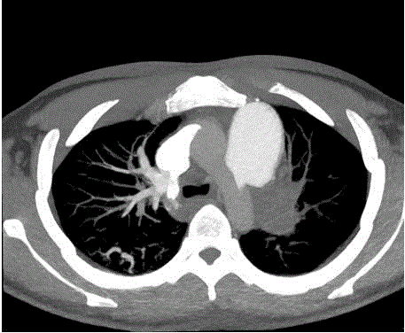
Figure 1
CTPA showed multiple PE, the left pulmonary artery was completely
embolized and the branches of right pulmonary artery were embolized.
Figure 2
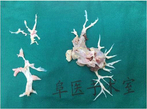
Figure 2
Excised pulmonary artery intima.
Discussion
This young patient got left chest pain at the onset of the disease.
The image results showed left inferior lobe pulmonary embolism
many times. Except smoking history, there weren’t certain risk
factors for venous thromboembolism (VTE) like fat, hyperlipidemia,
trauma, surgery etc, so some rare risk factors for thrombophilia
should be considered. From laboratory tests results we could see
LA, ACL, anti-β2GPI increased significantly and presented in high
titer after 4 months, from which APS could be diagnosed. There was no evidence exists to suggest other connective tissue diseases. So
PAPS should be considered as a diagnosis. The patient used novel
oral anticoagulant (NOVC) as standard regularly after developing
acute PE; but developed thromboembolic events twice again. Then
exercise intolerance, and hemoptysis were reported by the patient.
CTPA showed multiple pulmonary embolism. Several times of ECG
examination showed the increased size of the right ventricle and
right atrium and high systolic pressure of the pulmonary artery. The
diagnosis of CTEPH was made by his symptoms and imaging results.
Both venous and arterial thrombotic events can be presented
in APS patients. Deep vein thrombosis and PE are most common
thrombotic events. Most APS patient has more than one thrombus,
and it is common to see recurrent thrombotic events on APS patient.
According to other literature, the recurrence rate of thrombotic
events is up to 30% [3]. The rate of CTEPH developed from acute
PE ranges on 0.57%-4.7% from different studies. Some common
risk factors for this process include unexplained PE, recurrent PE,
big blood clot of PE, elderly, chronic inflammatory state, malignant
tumor, etc [4]. Some research indicates that aPLs are not only the
markers for APS, but also associated with the pathogen of CTEPH
[5]. Proposed mechanisms include that aPLs may inhibit the cascade
reactions catalyzed by phospholipids, which are also connected with
the development of CTEPH. Another hypothesis is that the formation
of microembolization in the course of APS makes a great contribution
to CTEPH [6]. According to the results of several cohort studies [7,8],
aPLs positive occurs in about 15-50% CTEPH patients. A high titer of
LA is involved with microembolization in PAPS [9]. The higher titer
of aPLS, the worse the prognosis is.
Effective, life-long anticoagulation is recommended for all
CTEPH patients. There is no evidence to prove NOVC is effective
as Warfarin [10]. In the early stage of disease, the patient didn’t get
a definite diagnose. He was considered as acute PE and only used
Rivaroxaban. Anticoagulant efficacy wasn’t evaluated regularly,
which caused the patient miss the optimal therapeutic time. While
going to the hospital, the patient experienced hemoptysis. The
CTPA showed pulmono-bronchial shunt of the left lung, which was
considered relative to CTEPH and was a difficult for anticoagulation.
Patients with CTEPH are more easily to get hemoptysis. That’s
because thrombus blocks both proximal and distal pulmonary artery,
and then led to the increase of blood supply provided by the bronchial
artery to pulmonary, and the increase of blood flow volume in the
capillary network between these two arteries. The capillary is easier
to rupture. So the anticoagulation for CTEPH patients should under
close surveillance to prevent bleed events.
Since pulmonary thromboendarterectomy (PTE) is the optimal
therapeutic schedule for CTEPH now, it is important to create
opportunity for operation. There is no clear evidence to prove that
patients with APS-CTEPH have fair prognosis after PTE. After PTE,
even though the patient still has triple antiphospholipid antibodies
(aPLs) positive; he felt obviously symptom relieve; and ECG showed
the recovery of cardiac structure and pulmonary arterial pressure on
the six month follow-up. From this case we can say that APS-CTEPH
is not a contraindication to PTE. After evaluate the risk of thrombosis
in an experienced treatment center and standard anticoagulation
treatment, patients with APS-CTEPH could accept PTE to get a good
prognosis.
References
- Miyakis S, Lockshin, Atsumi T, Branch DW, Brey RL, Cervera R, et al. International consensus statement on an update of the classification criteria for definite antiphospholipid syndrome (APS). J ThrombHaemost, 2006;4:295-306.
- Kim NH, Delcroix M, Jenkins DP, Richard C, Philippe D, Pavel J, et al. Chronic thromboembolic pulmonary hypertension. J Am Coll Cardiol. 2013; 62:D92-D99.
- Cervera R, Piette JC, Font J, Khamashta MA, Shoenfeld Y, Camps MT, et al. Antiphospholipid syndrome: clinical and immunologic manifestations and patterns of disease expression in a cohort of 1,000 patients. Arthritis Rheum. 2002;46:1019-1027.
- Delcroix M, Kerr K, Fedullo P, Chronic Thromboembolic Pulmonary Hypertension. Epidemiology and Risk Factors. Ann Am Thorac Soc. 2016;13:S201-S206.
- Porres-Aguilar M, Pena-Ruiz MA, Burgos JD, Porresmunoz M, Hughes HW. Chronic thromboembolic pulmonary hypertension as an uncommon presentation of primary antiphospholipid syndrome. J Natl Med Assoc. 2008;100:734-736.
- Zuily S, Wahl D. Pulmonary hypertension in antiphospholipid syndrome. Curr Rheumatol Rep. 2015;17:1-10.
- Levarge BL, Channick RN. Chronic thromboembolic pulmonary hypertension: evolution in management. Curr Opin Pulm Med. 2014;20:400-408.
- Wolf M, Boyer-Neumann C, Parent F, Eschwege V, Jaillet H, Meyer D, et al. Thrombotic risk factors in pulmonary hypertension. Eur Respir J. 2000; 15(2):395-399.
- Stojanovich L, Kontic M, Djokovic A, Ilijevski N, Stanisavljevic N, Marisavljevic D. Pulmonary events in antiphospholipid syndrome: influence of antiphospholipid antibody type and levels. Scand J Rheumatol. 2012;41:223-226.
- Nazzareno G, Humbert M, Jean-Luc V, Gibbs S, Lang I, Torbicki A, et al.2015 ESC/ERS Guidelines for the diagnosis and treatment of pulmonary hypertension[J]. Eur Heart J. 2016; 31:1219-1263.