Case Report
Fogging Effect in Strokes: Are we aware of it?
Huda Al-Jadiry1 and Ahmed Q Hasan2*
1Department of Radiology, University of Texas Medical Branch, USA
2Department of Emergency Medicine, Louisiana State University, USA
*Corresponding author: Ahmed Q Hasan, Department of Emergency Medicine, Louisiana State University, USA
Published: 15 Nov, 2017
Cite this article as: Al-Jadiry H, Hasan AQ. Fogging Effect
in Strokes: Are we aware of it?. Ann
Clin Case Rep. 2017; 2: 1465.
Abstract
Fogging effect is a transitory phenomenon of concealed cerebral infarction on computed tomography (CT). This effect is generally noticed within 2-3 weeks from the stroke onset.
Introduction
CT scan is the initial diagnostic imaging tool to demonstrate Ischemic strokes and ruling out/in hemorrhage. They usually demonstrate Hypo density on non-contrast CT scan. Iso-dense infarcts are misleading for radiologists and physicians as they might be interpreted as normal in spite of a clinically suspected stroke [1,2]. Fogging reported to be seen in about 50% of the ischemic strokes [3]. Knowledge of this effect is crucial to avoid misinterpretation or underestimation of this important finding and my guide further management especially if anticoagulants are being considered.
Discussion
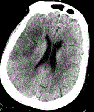
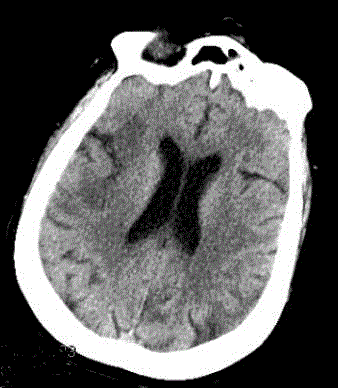
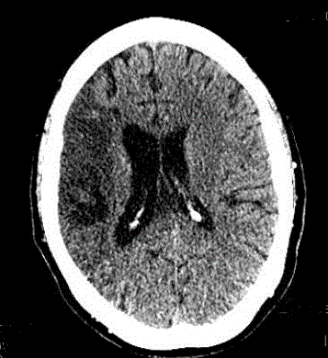
The fogging effect was first described in 1979 by Becker et al. [1]. It is defined as transient density equalization of the area of infarcted cerebral parenchyma in reference to normal parenchyma during the subacute phase of the infarct [2]. CT scan is the method of choice to monitor the evolution of infarcts. In the hyperacute phase, initial brain CT scans may be normal and is usually performed to exclude other potential causes of patients` symptoms. During the acute phase, the infarct will appear as an area of cortical hypodensity, blurring/loss of gray white matter differentiation, gyral swelling, and might causes mass effect (Figure 1). Subacute to chronic phase of the infarct usually demonstrates progressive lower attenuation till it becomes CSF density that reflects tissue death and necrosis [4] (Figure 2). The transient normalization of the CT scan during the subacute phase is attributed to migration of lipid-laden macrophages into the infarcted tissue, proliferation of the capillaries and redistribution of edema [5] (Figure 3). This phenomenon has been also described on T2 WI and presumed to be due the same pathophysiological process as it occurs in the same time frame [6]. Dekeyzer et al. [7] had described normalization of the immediate postinterventional CT scan. Given the short interval between pre and postinterventional CT Scan it is unlikely to be resulted from the afro mentioned physiological process. Dekeyzer et al. [7] self assumed it could be sequele of contrast leak due to disruption of blood brain barrier. Although this phenomenon has been described before, it remains a source of confusion for radiology residents, general radiologist and neurologist especially in the emergency setting and absence of prior infarct imaging. In conclusion, fogging of the subacute ischemic infarct is a temporary imaging phenomenon that masks a serious finding, which needs to be kept in mind when strokes are being worked up. Knowledge about this effect and awareness are of paramount importance as they minimize the risk of underestimating strokes and compromising patient care.
Figure 1
Figure 1
Initial non-contrasted CT scan of the brain, which demonstrates transcortical right parietal lobe infarct.
Figure 2
Figure 2
weeks later, non-contrasted CT of the head demonstrated fogging of right parietal lobe infarct.
Figure 3
References
- Becker H, Desch H, Hacker H, Pencz A. CT fogging effect with ischemic cerebral infarcts. Neuroradiology. 1979; 18: 185-192.
- Skriver EB, Olsen TS. Transient disappearance of cerebral infarcts on CT scan, the so-called fogging effect. Neuroradiology. 1981; 22: 61-65.
- O'Brien P, Sellar RJ, Wardlaw JM. Fogging on T2-weighted MR after acute ischaemic stroke: how often might this occur and what are the implications? Neuroradiology. 2004; 46: 635-641.
- Osborne, Salzman, Jhaveri. Diagnostic imaging Brain. Philadelphia: Elsevier. Section. 2016;4:358-362.
- Chalela JA, Kasner SE. The fogging effect. Neurology. 2000; 55: 315.
- Scuotto A, Cappabianca S, Melone MB, Puoti G. MRI "fogging" in cerebellar ischaemia: case report. Neuroradiology. 1997; 39: 785-787.
- Dekeyzer S, Reich A, Othman AE, Wiesmann M, Nikoubashman O. Infarct fogging on immediate post interventional CT-a not infrequent occurance. Diagnostic neuroradiology. 2017; 59: 853-859.


