Case Report
Intrathymic Mature Teratoma with Rupture into the Lung and Bronchus
Sani Rabiou1*, Marouane Lakranbi1, Yassine Ouadnouni1,2 and Mohamed Smahi1,2
1Department of Thoracic Surgery, University Hospital Hassan II, Morocco
2Department of Medicine and Pharmacy, Sidi Mohamed Ben Abdellah University, Morocco
*Corresponding author: Sani Rabiou, Department of Thoracic Surgery, University Hospital Hassan II, Fez, Morocco
Published: 12 Oct, 2017
Cite this article as: Rabiou S, Lakranbi M, Ouadnouni Y,
Smahi M. Intrathymic Mature Teratoma
with Rupture into the Lung and
Bronchus. Ann Clin Case Rep. 2017;
2: 1442.
Abstract
A young 19-year-old patient consults for a productive cough with purulent sputum with a notion of trichoptysis. The standard chest x-ray revealed an excavated excavation which sat at the level of the middle part of the left lung. The thoracic CT scan revealed a mediastinopulmonary cystic lesion containing area bubbles with numerous calcifications. The surgical gesture consisted of a complete resection of the lesion extended to the lingual. Anatomopathological examination of the surgical specimen confirmed the diagnosis of an intra-thymic teratoma containing cartilaginous, pancreatic, and osseous and sebaceous glands.
Keywords: Mature teratoma; Surgery; Complication
Introduction
Mature teratoma is the most common primary germ cell tumor in the mediastinum. These tumors rarely rupture into the adjacent structures, such as the pleural space, pericardium, lung parenchyma or tracheal tree. Thoracic CT - Scanis an examination in the diagnosis and therapeutic strategy. We report a case of mature mediastinal teratoma with rupture into the lung, and bronchus.
Case Presentation
A young 19-year-old patient consults for a productive cough with purulent expectorations evolving for 3 months in a febrile context. Interviewing revealed the notion of intermittent chest pain with an episode of trichoptysis (sputum containing hair). Clinical examination found a patient in very good general condition with a heart rate at 78 per minute and a respiratory rate at 21 cycles per minute. There was a clinical infectious syndrome with a fever at 38.5 °C and pleuropulmonary examination noted the presence of a decrease in the transmission of vesicular murmur of the left lung. The standard chest x-ray revealed an excavated opacity which sat at the level of the middle part of the left lung. Viewed the context of hydatid endemic in our country, the diagnosis of a pulmonary hydatid cyst ruptured in the bronchi had been evoked. But before the notion of trichoptysis with sputum bristles, a thoracic computed tomography was performed. It had revealed a mediastinopulmonary cystic lesion containing area bubbles with numerous calcifications (Figure 1A). Surgical exploration by postero-lateral thoracotomy had found a mediastinal cystic lesion containing sebum and hair, broken and fistulated in the left lung (Figure 1B). The gesture consisted of a complete resection of the lesion extended to the lingual. The postoperative sequences were marked by the occurrence of atelectasis requiring fibro aspiration with kinesia active respiratory therapy. The patient was discharged at D+6 postoperative after removal of the thoracic drain. Anatomopathological examination of the surgical specimen confirmed the diagnosis of an intrathymic teratoma containing cartilaginous, pancreatic, and osseous sebaceous glands.
Figure 1
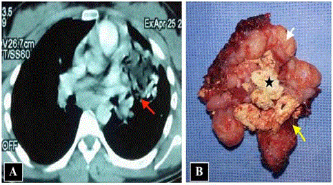
Figure 1
(A) CT scan showing a mediastinopulmonary cystic lesion,
ruptured, calcium and fatty density, containing area bubbles, indicative of
communication with the bronchi (red arrow). (B) Opening of the operating
room showing the presence of fat (white arrow), cartilaginous tissues, sebum
(black star) and hairs (yellow arrow).
Discussion
The rupture of a cystic mediastinal teratoma is rare but always symptomatic [1]. The mechanism of this rupture is still controversial, although ischemia, infection and chronic inflammation have been incriminated [1]. On the other hand, ectopic production of proteolytic or digestive enzymes favors inflammatory adhesion with erosion of adjacent structures [1]. Most authors have reported a rupture of a mediastinal teratoma following autolysis caused by the digestive enzymes released by the pancreatic tissue or salivary gland contained in the teratoma itself [1-3]. The presence of trichoptysis is pathognomonic of a rupture of a teratoma in the lung or in the bronchi. Thoracic computed tomography is the best paraclinic examination to study not only teratoma but also its relationship to adjacent structures. It also allows the planning of surgical strategy by seeking the presence of inflammatory adhesions between the teratoma and the neighboring organs. Cheung et al. [4] report the CT findings of a case of mediastinal cystic teratoma before and after rupture, and say that features of bursting of the spherical fatty component and intrapulmonary bronchial invasion are also suggestive of rupture of the mediastinal teratoma. The management of a ruptured teratoma is a surgery without delay. It confirms the diagnosis of the mature teratoma by eliminating the presence of an immature component and avoiding any complication due to its rupture, which can lead to haemoptysis, acute respiratory distress or cardiac tamponade [5]. This surgery is very haemorrhagic because of the adhesions between the mediastinal structures such as the phrenic nerve, the aorta, the vena cava and the pulmonary vessels [5]. Antibiotic treatment should be continued in order to avoid postoperative infectious complications.
Conclusion
The rupture or fistulization of a mature mediastinal teratoma reflects the delayed diagnosis. This implies further surgical intervention without delay to confirm the diagnosis by eliminating the presence of immature or malignant components, thus avoiding the occurrence of fatal complications.
References
- Choi SJ, Lee JS, Song KS, LimTH. Mediastinal teratoma: CT differentiation of ruptured and unruptured tumors.AJR Am J Roentgenol. 1998; 171: 591-594.
- Sasaka K, Kurihara Y,Nakajima Y, Seto Y, Endo I, Ishikawa T, et al. Spontaneous rupture: acomplication of benign mature teratomas of the mediastinum. AJR Am JRoentogenol. 1998; 170: 323-328.
- Serraj M, Lakranbi M,Ghalimi J, Ouadnouni Y, Smahi M. Mediastinal mature teratoma with complexrupture into the lung, bronchus and skin: a case report. World J Surg Oncol.2013;11:125.
- CheungYC, Ng SH, Wan YL, Pan KT. Ruptured mediastinal cystic teratoma withintrapulmonary bronchial invasion: CT demonstration. Br J Radiol. 2001; 74:1148-1149.
- Smahi M, Achir A, Chafik A, AlAziz AS, El Messlout A,Benosman A. Mature teratoma of the mediastinum. Ann Chir. 2000, 125: 965-971.