Case Report
A Bilateral Vertebrobasilar Stroke Mimicking a Bradycardia: A Case Report
Zidouh S*, Jidane S, Nabhani T, Chouaib N, Sirbou R, Belkouch A and Belyamani L
Department of Emergency and Critical Care, Military Hospital of Instruction, Mohammed V University Rabat,
Morocco
*Corresponding author: Saad Zidouh, Department of Emergency and Critical Care, Faculty of medicine and pharmacy, Military Hospital of Instruction, Mohammed V University Rabat, Agdal 10500 Rabat, Morocco
Published: 18 Jul, 2017
Cite this article as: Zidouh S, Jidane S, Nabhani T,
Chouaib N, Sirbou R, Belkouch A, et
al. A Bilateral Vertebrobasilar Stroke
Mimicking a Bradycardia: A Case
Report. Ann Clin Case Rep. 2017; 2:
1403.
Abstract
We report the case of a man admitted to the emergency department for a stroke with an unusual presentation: bradycardia due to Atrial Fibrilation with slow ventricular response. The cerebral CT scan showed a bilateral vertebro-basilar acute ischemic stroke. The evolution was marked by neurological degradation with a Glasgow Coma Scale at 6/15. The patient was admitted to the intensive care unit and intubated. Three days later, he presented a massive hemorrhagic infarction and died shortly after.
Keywords: Bradycardia; Vertebrobasilar- ischemic stroke
Case Presentation
We report the case of a 79-year-old man, known to be hypertensive and coronary, admitted to
the emergency department for confusion and lipothymia since 4 hours, without fever. The Glasgow
Coma Scale (GCS) is at 13/15 (E4, V4, M5). The patient wasn’t on anticoagulants or antiplatelet
agents. The neurological examination revealed no anomalies.
Shortly after his admission to the vital emergency room (VER), he presented a bradycardia,
his heart rate decreased from 97 to 32 bpm, associated toan arterial hypotension at 73/41 mmHg.
A concomitant electrocardiogram with the rhythm disorder objectified an atrial brady-fibrillation.
The patient recovered quickly after a bolus of Atropine. Indeed, the heart rate increased to 85 bpm
and blood pressure to 133/54 mmHg.
No biological abnormalities were found, in particular an ultrasensitive troponin, which was
negative. An echocardiographic supplement found global dyskinesia, with septal akinesia and left
atrial dilatation. The evolution was marked by neurological degradation, with a GCS at 6/15, which
motivated an orotracheal intubation to protect the airways. A cerebral CT scan without injection
was made, and objectified a bilateral vertebrobasilar ischemic stroke associated to a vascular
leucoencephalopathy (Figure 1).
The patient was transferred to the intensive care unit and kept for 48 hours in sedation for
cerebral detente with preventive treatment of intracranial hypertension. A control cerebral CT scan
on the third day showed a massive hemorrhagic infarction with edema and mass effect compressing
the fourth ventricles. The patient died few days later.
Figure 1
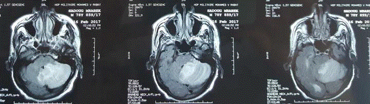
Figure 1
MRI images showed a bilateral vertebrobasilar ischemic stroke.
Discussion
Among all cases of stroke, 80% are ischemic, and 25% of infarcts are located in the vertebrobasilar arterial territory [1]. Posterior circulation stroke can have diverse presentations that differ from strokes in anterior circulation in relation to etiology, clinical features,
and prognosis. Posterior circulation stroke can be presented with
vertigo, ataxia, vomiting, headache, cranial nerve abnormalities,
bilateral long tract neurological sign, ‘‘locked in’’ syndrome or
impaired consciousness, and complex ocular signs or cortical
blindness with high mortality and morbidity [1,2].
Overall, the most common findings were bulbar and
pseudobulbar signs (73.6%), weakness (56.3%), vertigo or dizziness
(54.0%), ataxia (48.75%) that is due to the involvement of cerebellum
or its connections [2]. The prospective study of Mehndiratta showed
vomiting in 41% and headache in 31% of cases [3]. In our case the
initial symptomatology was poor, and only confusion has marked the
clinical picture.
Patients with acute ischemic stroke can develop stress-induced
cardiomyopathy, clinically manifesting as acute coronary syndrome
reconsidering unnecessary coronary interventions [4], this maybe
explain the bradycardia developed shortly after the admission in
our case. It has been tried to understand the complex, yet mostly
unknown, regulatory relationship between the heart and the brain
and damage in brain in any form can impede normal cardiovascular
function [5].
About risk factors, Veotsch objective in her medical registry that
hypertension was the most frequent risk factor (66.7%), followed by
hyperlipidemia (37.9%) and coronary artery disease (33.3%) [2]. Our
patient had as cardiovascular risk factor in addition to the male sex,
hypertension and coronary artery disease.
When considering clinical symptoms and signs, decreased
level of consciousness, tetraparesis or tetraplegia, and pupillary
abnormalities were significantly associated with worse outcome. The
mortality rate was at 2.3% [2]. In our case the death was caused by a
common complication of ischemic stroke, illustrated by hemorrhagic
infarction.
References
- Campanholo KR, Conforto AB, Rimkus CM , Miotto EC. Cognitive and Functional Impairment in Stroke Survivors with Basilar Artery Occlusive Disease. Behav Neurol. 2015; 2015: 7.
- VoetschB, DeWitt LD, Pessin MS, Caplan LR. Basilar Artery Occlusive Disease in the New England Medical Center Posterior Circulation Registry. Arch Neurol. 2004; 61: 496-504
- Mehndiratta M, Pandey S, Nayak R, andAlam A. Posterior Circulation Ischemic Stroke Clinical Characteristics, Risk Factors, and Subtypes in a North Indian Population: A Prospective Study. The Neurohospitalist. 2012; 2: 46-50.
- Lee JS, Pyung Chun Oh PC, Koh KK. Acute ischemic stroke mimicking acute coronary syndrome. Int J Cardiol. 2016; 221: 560-561.
- Bybee KA, A Prasad. Stress-related cardiomyopathy syndromes. Circulation. 2008; 118: 397-409.