Clinical Image
Angina Associated with Dynamic Right Ventricular Compression of Anomalous Left Main Coronary Artery
Szolnoky J#, Eichinger S*# and Eichinger WB
Department of Cardiac Surgery, Hospital Bogenhausen, Germany
#Both the authors contributed equally
*Corresponding author: Simone Eichinger, Department of Cardiac Surgery, Hospital Bogenhausen, Klinikum Bogenhausen, Englschalkingerstrasse 77, 81925 Munich, Germany
Published: 12 Jul, 2017
Cite this article as: Szolnoky J, Eichinger S, Eichinger WB.
Angina Associated with Dynamic Right
Ventricular Compression of Anomalous
Left Main Coronary Artery. Ann Clin
Case Rep. 2017; 2: 1400.
Abstract
Left main coronary artery arising from the right anterior sinus with anomalous course may
predispose to myocardial ischemia, infarction or sudden death. This coronary anomaly can be
divided in various subtypes, with one of them being a very rare anatomic variation where the left
main coronary artery is located anterior to the right ventricular outflow tract [1].
We report a 60-year-old patient, who was admitted with stable exercise induced angina refracter
to standard medication. Diagnostic coronary angiography revealed a coronary anomaly with origin
of the left main coronary artery from the right coronary sinus and anterior course proximal to the
pulmonary trunk with severe dynamic compression of the vessel (Figure 1). The patient was referred to bypass surgery. Surgery was performed with extracorporal circulation. During cardioplegic arrest
the aortic and pulmonary root was examined, and a right ventricular intramuscular course of the
anomalous left main coronary proximal to the pulmonary trunk was found. The vessel was carefully
dissected from the ventricle muscle to dissolve its dynamic muscular compression (Figure 2). Four 5.0 sutures were used to fixate the myocardiac muscle tissue in order to avoid recurring compression
of the left main coroanry artery.
On the fifth postoperative day a 64-slice computed tomography
showed no residual systolic compression of the left main coronary
artery (Figure 3). After uneventful postoperativ course the patient
was discharged one week postsurgically.
Figure 1
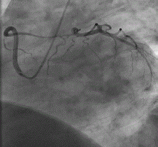
Figure 1
Diagnostic coronary angiography.
Figure 2
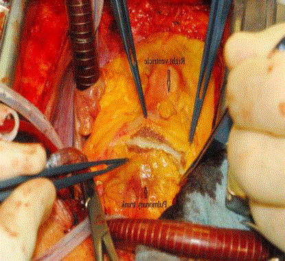
Figure 2
Dynamic muscular compression.
Figure 3
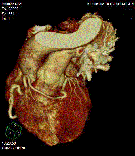
Figure 3
Computed Tomography showed no residual systolic compression
of the left main coronary artery.