Case Report
Giant Chylolymphatic Mesenteric Cysts in Children: A Preoperative Enigma
Lone AY, Randhawa JS, Samujh R*, Dash V and Rao SG
Department of Pediatric Surgery, Postgraduate Institute of Medical Education and Research (Pgimer), India
*Corresponding author: Samujh R, Department of Pediatric Surgery, Postgraduate Institute of Medical Education and Research (Pgimer), Chandigarh, India
Published: 12 Jun, 2017
Cite this article as: Lone AY, Randhawa JS, Samujh R,
Dash V, Rao SG. Giant Chylolymphatic
Mesenteric Cysts in Children: A
Preoperative Enigma. Ann Clin Case
Rep. 2017; 2: 1373.
Abstract
Although mesenteric cysts in general have been reported in the literature fairly frequently, chylolymphatic cysts in the pediatric age group are extremely rare in the modern medical literature. Ultrasonography and computed tomography suggest the diagnosis but the picture is not clear until demonstrated intraoperatively. Intra-operatively, similar findings can be seen in cystic lymphangioma, retroperitoneal cystic teratoma, caseating tubercular lymph nodes, and hydatid cysts. Even lymphoma and duplication cysts may also give similar appearances. Excision biopsy is then recommended to differentiate these cases. Histopathology is confirmatory and differentiates chylolymphatic cysts from all these lesions. We present our experience with two such cases that presented to our center.
Keywords: Chylolymphatic; Mesenteric cyst; Ileum; Abdominal pain
Introduction
A chylolymphatic cyst is a rare variant of a mesenteric cyst [1,2]. These cysts present within the mesentery, lined with a thin endothelium or mesothelium and filled with chylous and lymphatic fluid [3]. Although mesenteric cysts in general have been reported in the literature fairly frequently, chylolymphatic cysts in the pediatric age group are extremely rare in the modern medical literature [2], therefore very little information is available regarding their presentation which can vary from asymptomatic to an acute abdomen with intestinal obstruction. Ultrasonography and computed tomography can provide an idea regarding the diagnosis but the intraoperative findings are often quite different. Histopathological examination of the specimen is required for confirmation. Complete excision of the cyst yields excellent result. In an attempt to reinforce the diagnostic and treatment strategy, we present our experience with two such cases that presented to our center within a short span of two weeks.
Case Presentation
Case 1
A 4 years old female child presented with abdominal pain of 6 months duration. The pain
was dull, aching and continuous in nature. It was associated with mild abdominal distension
noted for the last 1 month. The general physical examination was unremarkable. The abdomen
was mildly distended but soft and suggestive of diffuse ascites rather than a discrete palpable mass.
An ultrasonography (USG) and computed tomography scan (CT scan) of the abdomen revealed a
large, septated, cystic mass with hypodense fluid attenuation. The biochemical parameters including
serum amylase and lipase were within normal limits. With a preoperative suspicion of mesenteric
cyst or pelvic cyst, the patient was posted electively for exploratory laparotomy but had to be taken
up in the emergency because of a 10 days history of increasing abdominal pain and decreased oral
intake. There was no history of vomiting or constipation. Upon surgical exploration, there was a
huge cystic mass about 15cm by 18cm in dimension, filled with white milky fluid suggestive of a
chylolymphatic cyst, occupying the mesentery of proximal ileum (Figure 1). The cyst was enucleated from the mesentery and excised completely along with 5cm of ileum closely related to it (Figure 2). An ileoileal end to end anastomosis was done. Feeds were started from 5th postoperative day
and the patient was discharged on 7th postoperative day without any postoperative complications.
The histopathological examination of resected specimen confirmed the diagnosis of chylolymphatic
mesenteric cyst. At 6 months of follow up the patient is feeding adequately and doing well.
Case 2
A 5 years old male child presented with history of abdominal pain for the last 3 years. The pain was continuous, dull, aching but intermittently increased in intensity
and the child needed analgesics for the relief. The pain increased in
intensity and in frequency over the last 2 months. The patient was
investigated with USG and CT scan of abdomen suggesting a huge
intraperitoneal cystic mass and posted electively for exploration
(Figure 3). This patient also underwent emergency exploratory
laparotomy due to features of acute intestinal obstruction. Upon
surgical exploration, there was a huge 10cm by 15 cm cystic mass
filled with white milky fluid in the mesentery of jejunum 35 cms
from duodenojejunal flexure (Figure 4). Resection of about 4cm of
jejunum with end to end anastomosis was needed in this case also
with uneventful postoperative course. The patient was discharged by
7th postoperative day and is doing well after 6 months of follow up.
The histopathology confirmed a chylolymphatic mesenteric cyst.
Figure 1
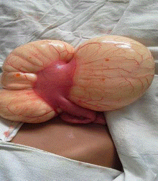
Figure 1
Intraoperative picture – Case 1- Huge chylolymphatic mesenteric
cyst attached to mesentery of ileum.
Figure 2
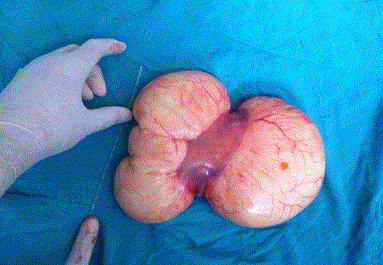
Figure 2
Resected specimen – Case 1.
Figure 3
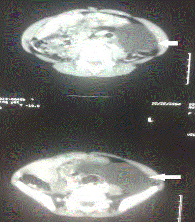
Figure 3
CT scan-Arrows pointing at huge hypodense fluid attenuation of
chylolymphatic mesenteric cyst– Case 2.
Figure 4
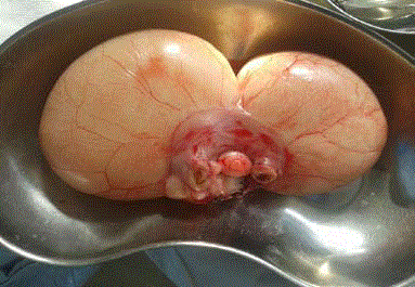
Figure 4
Resected specimen – Case 2.
Discussion
Chylous cysts are rare variants of mesenteric lesions and
constitute 7.3% to 9.5% of all abdominal cysts [1]. There are very few
cases of pediatric chylolymphatic cysts reported in the literature. The
clinical presentation is varied ranging from asymptomatic to palpable
abdominal mass, diffuse abdominal fullness, abdominal pain and in
extreme cases an acute abdomen with features of intestinal obstruction.
These two cases presented here stand testimony to the fact that a
mesenteric cyst with torsion, hemorrhage or intestinal obstruction
can present as an acute abdomen and should be kept in mind as one of
the differential diagnosis. Beahrs et al. [4] classified mesenteric cysts
into four groups based on etiology: embryonic or developmental; traumatic or acquired; neoplastic; and infective or degenerative.
Recently, a pathologic classification system has been proposed [5].
Type 1 (pedicled) and Type 2 (sessile) are limited to the mesentery and
can be excised completely with or without resection of the involved
gut. Type3 and Type4 (multicentric) extend into the retroperitoneum
and require complex operations and often sclerotherapy as well.
Based on the contents of the cyst, the mesenteric cyst can be divided
into serous, chylous, hemorrhagic and chylolymphatic cyst. The
chylolymphatic cyst, as indicated by its name, contains both chyle
and lymph. The accumulation of chyle and lymph is considered
to be the result of an imbalance between the inflow and outflow of
fluid [1]. Radiological investigations form an integral part of the
management of these lesions. A plain abdominal radiograph may
show a gasless, homogenous mass defect displacing the bowel loops
around it. In a child with an obstructed intestine, multiple air-fluid
levels will be seen on an erect abdominal radiograph. Barium studies
are now only of historical interest; abdominal ultrasonography is
currently the imaging procedure of choice. This delineates the nature
of the mass, organ or site of the origin, and the extent and associated
mass effects on the kidney or liver, if any. Computed tomography
adds little additional information; however, contrast-enhanced film
can show the relationship of the bowel and other vital structures
to the lesion. Some authors have described the characteristic
appearance of a chylolymphatic cyst on computed tomography and ultrasonography in the form of the presence of fluid levels of differing echodensities, that is to say, an upper fatty echodensity of chyle on
top of the water echodensity of lymph in a well-defined cystic lesion
[6,7]. The different surgical approaches used are marsupialization,
sclerotherapy, drainage, enucleation, percutaneous aspiration, and
excision of the cyst with or without resection of the involved gut [8,9].
Due to high recurrence rates associated with marsupialization and
drainage, complete excision of the cyst should be attempted whenever
possible [4]. In adults, the cyst can often be enucleated or 'shelled
out' from between the leaves of the mesentery, however, in children,
bowel resection is frequently required [9]. Literature mentions
instances where laparoscopic removal of mesenteric cysts has been
tried successfully, but this might have been difficult in our patients,
especially with large sized chylolymphatic mesenteric cysts [10].
Intra-operatively, similar findings can be seen in cystic
lymphangioma, retroperitoneal cystic teratoma, caseating tubercular
lymph nodes, and hydatid cysts. Even lymphoma and duplication cysts
may also give similar appearances. Excision biopsy is recommended
to differentiate these cases. Histopathology is confirmatory and
differentiates chylolymphatic cysts from all these lesions. Cystic
lymphangioma has a striking resemblance to chylolymphatic
mesenteric cysts both grossly and microscopically. Some authors
consider chylolymphatic mesenteric cysts to be a type of cystic
lymphangioma, but the medical literature also shows some authors
describing chylolymphatic cysts as a variant of mesenteric cysts [3,5].
The absence of smooth muscle and lymphatic spaces in the wall of
the cyst differentiates mesenteric cysts from cystic lymphangioma [3].
Conclusion
Although very rare, chylolymphatic mesenteric cysts should be kept in mind as one of the differential diagnosis of cystic masses in children and can have a varied presentation ranging from asymptomatic to abdominal pain, mass or an acute abdomen with features of intestinal obstruction.
References
- Engel S, Clagett OT, Harrison EG Jr. Chylous cysts of the abdomen. Surgery. 1961; 50: 593-599.
- Gupta AR, Nanavati RN, Fernandez AR, Kalgutkar A, Nathani R, Deshmukh SS. Chylous mesenteric cyst: an unusual cause of neonatal intestinal obstruction. Indian Pediatr. 1992; 29: 511-513.
- Takiff H, Calabria R, Yin L, Stabile BE. Mesenteric cysts and intraabdominal cystic lymphangiomas. Arch Surg. 1985; 120: 1266-1269.
- Beahrs OH, Judd ES Jr, Dockerty MB. Chylous cysts of the abdomen. Surg Clin North Am. 1950; 30: 1081-1096.
- Losanoff JE, Richman BW, El-Sherif A, Rider KD, Jones JW. Mesenteric cystic lymphangioma. J Am Coll Surg. 2003; 196: 598-603.
- Fujita N, Noda Y, Kobayashi G, Kimura K, Watanabe H, Masu K, et al. Chylous cyst of the mesentery: US and CT diagnosis. Abdom Imaging. 1995; 20: 259-261.
- Phillips GW, Senapati A, Young AE. Chylolymphatic mesenteric cyst: a diagnostic appearance on computer tomography. Br J Radiol. 1988; 61: 413-414.
- Hebra A, Brown MF, McGeehin KM, Ross AJ. Mesenteric, omental, and retroperitoneal cysts in children: a clinical study of 22 cases. South Med J. 1993; 86: 173-176.
- Kurtz RJ, Heimann TM, Holt J, Beck AR. Mesenteric and retroperitoneal cysts. Ann Surg. 1986; 203: 109-112.
- Raghupathy RK, Krishnamurthy P, Rajamani G, Babuji N, Diriviraj R, Mohan NV, et al. Intraabdominal cystic swelling in children - laparoscopic approach, our experience. J Indian Assoc Pediatr Surg. 2003; 8: 213-217.