Case Report
Unsuspected Foreign Body in Parotid Gland as a Cause of Facial Palsy: A Rare Case Report
Abhijeet Singh, Anand S, Gyan Ranjan Nayak and Naresh Panda*
Department of Otolaryngology Head and Neck Surgery, Post Graduate Institute of Medical Education and
Research, India
*Corresponding author: Naresh Panda, Department of Otolaryngology Head and Neck Surgery, Post Graduate Institute of Medical Education and Research, Chandigarh, India
Published: 12 Apr, 2017
Cite this article as: Singh A, Anand S, Nayak GR, Panda
N. Unsuspected Foreign Body in
Parotid Gland as a Cause of Facial
Palsy: A Rare Case Report. Ann Clin
Case Rep. 2017; 2: 1334.
Abstract
Parotid gland injuries are often associated with a number of sequelae. Assault and accidents contribute to nearly 90% of the parotid gland injuries. We report a case of an 18 year old male who presented with a history of trauma to the left side of face six weeks back, which he sustained after a wooden log fell from a height of about 10 feet. Patient developed delayed onset weakness over the lower part of face after three weeks of trauma. CT scan revealed the possibility of a foreign body within the sinus tract.
Keywords: Parotid foreign body; Discharging sinus; Organic foreign body; Parotid sinus
Introduction
Parotid gland injuries are often associated with a number of sequelae. Assault and accidents
contribute to nearly 90% of the parotid gland injuries. Major sequelae following injury include
sialoceles and fistulae. Conservative management is sufficient is more than 50% of the cases and
rest of the cases require surgical drainage [1]. Stenson’s duct when accessible should be repaired
primarily. Conservative management options include the use of antisialogogues, elastic bandages,
and refrain from oral intake until the injury is healed [2].
Very few cases require parotidectomy [3]. A thorough knowledge of the structure and function
of the parotid region is essential in management of these injuries. We report a case of an 18-year
old malewitha discharging sinus and swelling in the left parotid region with ipsilater allower motor
neuron facial palsy who failed to respond to conservative management.
Case Presentation
We report a case of an18 year old male patient who presented to our outpatient department with
a history of trauma to the left side of face six weeks back, which he sustained after a wooden log fell
from a height of about 10 feet. At that time patient had a small laceration over the left parotid region.
A small piece of wood was removed from the wound site by a physician at a primary care Centre
and started on oral antibiotics. Two weeks later patient started having purulent discharge from the
wound (Figure 1). Patient developed delayed onset weakness over the lower part of face after three
weeks of trauma for which a magnetic resonance imaging (MRI) was ordered by the physician and
subsequently referred to our Centre. Clinical examination revealed purulent discharge from the
sinus with diffuse induration over left parotid region, which was extending into left side neck as
well. Patient had House-Brackmann Grade III lower motor neuron facial palsy involving the buccal
and marginal mandibular division of facial nerve on the left side (Figure 2). Oral cavity, neck and
otological examination were unremarkable. MRI revealed a linear fluid distended sinus tract in left
intra parotid region involving the deep and superficial lobe of parotid reaching up to skin surface
(Figure 3). A contrast enhanced computed tomography scan (CT) was done after discussing MRI
with the radiologist. CT scan revealed the possibility of a foreign body within the sinus tract (Figure 4).
Patient was then taken up for surgical exploration under general anesthesia. Modified Blair’s
incision was made including the sinus tract. Intra-operatively, a piece of wood was found embedded
in the parotid parenchyma and was removed into (Figure 5). Cavity was thoroughly irrigated with
diluted antibiotic solution and wound was closed over a corrugated rubber drain. Patient made an
uneventful recovery and was discharged on oral antibiotics after five days (Figure 6). At four week
follow up facial palsy had recovered completely (House Brackmann Grade I) (Figure 7).
Figure 1
Figure 1
Discharging sinus in left parotid region.
Figure 2
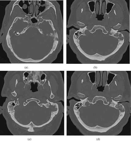
Figure 2
Left Grade III LMN facial palsy.
Figure 3
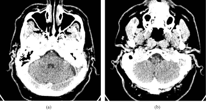
Figure 3
Left Grade III LMN facial palsy.
Figure 4
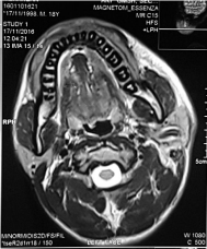
Figure 4
MRI revealed a linear fluid distended sinus tract in left parotid
region.
Figure 5
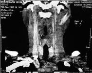
Figure 5
CECT Neck - Foreign body in left parotid region (Coronal).
Discussion
Injuries to salivary glands remain uncommon, with the number
one cause being penetrating trauma. Other causes of acute injuries
include blunt trauma and blast injuries. Salivary gland injuries are
relatively uncommon, only a few large series exist. A great deal of
experience comes from injuries sustained during World War II [4] (Figure 8).
Optimal outcome requires early recognition with an adequate
evaluation, directing proper management. Athorough history must
be obtained whenever possible. Important questions include timing
and nature of injury as well as other injuries. Valuable information
can be gathered by determining if the patient has been able to eat after sustaining the injury and the effect this has on the salivary
tissue. Additionally, if there has been drainage from the wound, the
character of this drainage can be suggestive. Examination requires
inspection of the gland and surrounding structures while comparing
it with the contralateral side for symmetry. Surrounding structures
include overlying skin, oral mucosa, and dental structures and
complete otological examination is indicated. Electroneurography
and electromyography represent a more sophisticated means of evaluation of facial nerve trauma. If degeneration of the facial nerve
exceeds 90% on electroneurography and it is within a period of three
weeks after the injury, surgical exploration may be indicated. After the
three-week period, electromyography may be indicated to determine
if a nerve is recovering [5] (Figure 9).
Magnetic resonance imaging is the gold standard imaging tool
for parotid neoplasms and parotid diseases. It is not necessarily the imaging study of choice for trauma to the parotid gland. Both CT
coronal and axial provide the best evaluation of the bony structure.
Additionally, CT can give excellent soft tissue delineation.
Penetrating injury to the parotid gland, in addition to involving
the parenchyma of the gland, also can involve Stensen's duct or the
facial nerve. The anterior border of the master muscle is an important
land- mark because lacerations posterior to this can injure the duct.
Although a superficial or total parotidectomy would eliminate the
salivary source, the morbidity in such a compromised bed may
outweigh its benefits. When identified, injuries to Stensen's duct
should be primarily repaired. Missing a duct injury may lead to the
development of a posttraumatic fistula or sialocele.
Figure 6
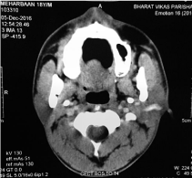
Figure 6
CECT Neck - Foreign body in left parotid region (Coronal).
Figure 7
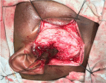
Figure 7
Intra operative.
Figure 8
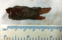
Figure 8
Wooden foreign body.
Figure 9
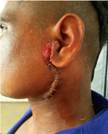
Figure 9
Postoperative day 5.
Conclusion
Post traumatic chronic discharging sinus along with facial palsy should be evaluated with a high index of suspicion for retained foreign body. Evaluation with appropriate imaging may guide towards proper surgical planning.
References
- Neilan RE, Kutz JW Jr. Langerhans cell histiocytosis of the temporal bone. Otol Neurotol. 2012;33(4):e31-2.
- Howarth DM, Gilchrist GS, Mullan BP, Wiseman GA, Edmonson JH, Schomberg PJ. Langerhans cell histiocytosis: diagnosis, natural history, management, and outcome. Cancer. 1999;85(10):2278-90.
- Blumberg JM, Malhotra A, Wu X, Virk RK, Kveton JF, Michaelides EM. Langerhans Cell Histiocytosis of the Temporal Bone with Otic Capsule Involvement. Clin Neuroradiol. 2015.
- Modest MC, Garcia JJ, Arndt CS, Carlson ML. Langerhans cell histiocytosis of the temporal bone: A review of 29 cases at a single center. Laryngoscope. 2016;126(8):1899-904.
- Yildirim-Baylan M, Cureoglu S, Paparella MM. Langerhans' cell histiocytosis of the temporal bone. Otol Neurotol. 2012;33(2):e15-6.
- Coleman MA, Matsumoto J, Carr CM, Eckel LJ, Nageswara Rao AA. Bilateral temporal bone langerhans cell histiocytosis: radiologic pearls. Open Neuroimag J. 2013;7:53-7.
- Wirtschafter JD, Nesbit M, Anderson P, McClain K. Intralesional methylprednisolone for Langerhans' cell histiocytosis of the orbit and cranium. J Pediatr Ophthalmol Strabismus. 1987;24(4):194-7.
- Abdel-Aziz M, Rashed M, Khalifa B, Talaat A, Nassar A. Eosinophilic granuloma of the temporal bone in children. J Craniofac Surg. 2014;25(3):1076-8.
- Satter EK, High WA. Langerhans cell histiocytosis: a review of the current recommendations of the Histiocyte Society. Pediatr Dermatol. 2008;25(3):291-5.
- Sweet RM, Kornblut AD, Hyams VJ. Eosinophilic granuloma in the temporal bone. Laryngoscope. 1979; 89 (10):1545-52.