Case Report
Temporal Bone Lesion: A Recurrence of Mandibular Langerhans Cell Histiocytosis
Abi-Akl P1, Haddad G1 and Zaytoun G1*
Department of Otolaryngology-Head & Neck Surgery, American University of Beirut Medical Center– Beirut-
Lebanon
*Corresponding author: Georges Zaytoun, Department of Otolaryngology-Head & Neck Surgery, American University of Beirut Medical Center– Beirut-Lebanon
Published: 12 Apr, 2017
Cite this article as: Abi-Akl P, Haddad G, Zaytoun G.
Temporal Bone Lesion: A Recurrence
of Mandibular Langerhans Cell
Histiocytosis. Ann Clin Case Rep. 2017;
2: 1330.
Abstract
Background: Langerhans Cell Histiocytosis results from the abnormal clonal proliferation of
Langerhans cells. In adults, it most commonly presents in the form of Eosinophilic Granuloma, a
focal osteolytic lesion.
Case Report: A 51-year-old man with a history of cured Eosinophilic Granuloma of the mandible
presented with otologic complaints. His physical examination was normal, but further imaging
showed a temporal bone lesion, which was found to be a recurrence of the Langerhans Cell
Histiocytosis.
Conclusion: Although the recurrence of eosinophilic granuloma is very rare, it is important to rule
it out even in the context of a normal physical examination.
Introduction
Under normal physiological conditions, Langerhans cells are antigen presenting cells located in the dermis and that migrate to lymph nodes to activate the immune system when needed [1]. Langerhans Cell Histiocytosis (LCH) is by definition a clonal proliferation of Langerhans cells outside the dermis that damage and erode surrounding tissues in which they accumulate [2,3]. The pathophysiology of LCH is still ambiguous with theories of neoplastic, inflammatory, infectious and genetic etiologies quoted in the literature [1,3]. The incidence of LCH in the general population ranges from 1 to 9 cases per million per year [1-3]. It has a predilection for males with a male to female ratio of 1.7:1. The peak age of incidence is 1-4 years [3]. The disease severity of LCH spans a spectrum ranging from a unifocal disease affecting only one system, to a life-threatening, multisystem condition [1,2]. It is broadly sorted into 3 categories: Eosinophilic Granuloma, Hand- Schuller-Christian disease and Letterer-Siwe disease. Eosinophilic Granuloma is characterized by a unifocal osteolytic lesion with rare systemic symptoms. Hand-Schuller-Christian disease presents with multifocal osteolytic lesions in young children, usually with no extra-skeletal manifestations. Letterer-Siwe disease carries the worst prognosis of the three and presents in children less than 3 years of age, with multi-organ involvement. Mortality usually occurs due to multi-organ failure at a rate of 50% of cases [1,4]. Otologic manifestations of LCH occur in 8-61% of cases, with a temporal bone mass being the most common one. Bilateral involvement is found in one-third of patients with temporal bone LCH. Diagnostic procedures include the history and physical exam, which most commonly reveal otorrhea and subjective hearing loss [3-5]. Biopsy of the temporal bone lesion is necessary for histopathologic confirmation. Computer Axial Tomography (CAT) scan remains the undisputed imaging modality of choice to diagnose temporal bone LCH and its bony involvement [1,4-6]. Several options are discussed for the treatment of LCH [5]. Surgical excision and/or curettage are usually only reserved for local, confined disease, while chemotherapy is the treatment of choice especially for disseminated disease, for multi-organ involvement and for recurrence [1,4]. There are reports of successful intra-lesional steroid infiltration [7]. Radiotherapy is also mentioned in the literature and is most commonly reserved for recurrent disease [1,4]. Several authors however recommend a combination of the methods mentioned above to optimize cure [2,8]. Prognosis of LCH depends on the nature of the disease. Relapse and death occur infrequently in localized unifocal disease while multisystem disease is associated with grimmer outcomes [3].
Case Presentation
Mr. N. A. is a 51-year-old white male who presented in August 2016 with subjective hearing loss and tinnitus in the left ear of 10 days duration. He also reported a brief episode of bilateral hearing loss and left otalgia 2 months prior to presentation that had resolved spontaneously (Figure 1). He gave a history of histopathologically confirmed Langerhans Histiocytosis of the mandible in 2013, treated with curettage and local radiotherapy. Physical exam revealed normal ear canals bilaterally. There was no evidence of otitis externa or media. The tympanic membranes were intact bilaterally, with no evidence of middle ear effusion. The Rinne test was positive bilaterally, and the Weber test did not lateralize. There was no erythema or swelling around the mastoid area. Further evaluation revealed bilateral mild sensorineural hearing loss on audiogram. A CAT scan of the head was obtained and revealed large, irregular lytic changes and erosions involving most of the mastoid air cells and surrounding bones, communication with the surrounding deep soft tissues and with the intracranial cavity at the level of lateral border of the posterior cranial fossa (Figures 2 & 3). The erosion was in close proximity to the posterior border of the left temporomandibular joint. The soft tissue window cuts of the CAT scan showed partial extension of the tumor into attic of the left middle ear cavity and into the antrum. Biopsy of the left mastoid was performed and revealed Langerhans cell disease of the eosinophilic granuloma subtype. This was followed by a PET-CT scan, which showed minimal activity associated with left mastoid lesion, but otherwise no dissemination of the disease.
Figure 1
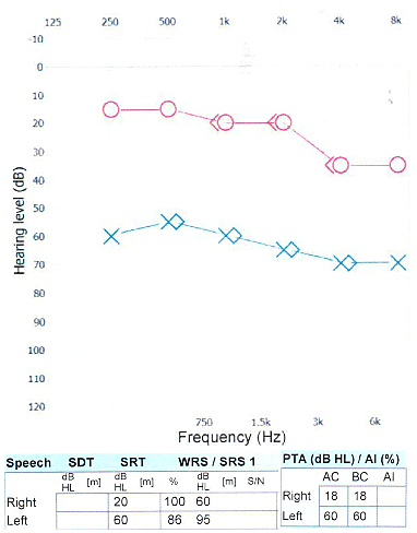
Figure 1
Audiogram done 2 months prior to presentation that reveals mild
right and moderately severe left sensorineural hearing loss.
Figure 2
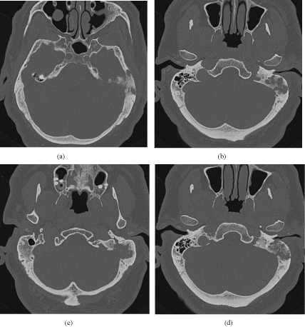
Figure 2
CAT scan cuts showcasing the left temporal bone mass. (a) Shows
lytic bony erosions of the left temporal bone. (b) Shows the obliterated mastoid
air cells on the left as compared to the right side. (c,d) show intracranial
extension at the level of the lateral wall of the posterior cranial fossa.
Figure 3
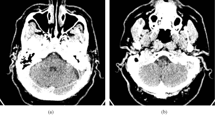
Figure 3
CT scan cuts showing the soft tissue window and extension of the
mass to the posterior cranial fossa.
Discussion
All the elements in our patient’s presentation are consistent the clinical picture of LCH as is evident in the literature. Indeed, LCH has more predilections for males who present with the most common otologic manifestation, a mass in the temporal bone [3- 5]. In addition, our patient complained of subjective hearing loss, confirmed by his pure tone audiogram, which is another common otologic manifestation of LCH [3-5]. Particular care was taken to rule out bilateral disease which may occur in roughly 30% of cases that present with a temporal bone mass [3-5]. The LCH in our patient falls under the category of Eosinophilic Granuloma since it is unifocal. Unifocal lesions in LCH are most common in the adult population, with an occurrence rate of 69-72% [9]. Our patient also belongs to the rare cases that suffer a relapse of monostotic EG (10% of cases) [3]. It is certainly peculiar that no abnormal findings were seen on physical exam. Both ears had normal external canals with no otorrhea and no evidence of tympanic membrane or middle ear pathology. Further imaging was performed merely because of a previous history of LCH in the setting of an otherwise unimpressive clinical examination. It is thus always important to have a high index of suspicion whenever there is a past history of LCH, even if the physical examination is unrevealing. Back in 2013 when he had first presented with LCH of the mandible, the patient was treated with surgical curettage and radiotherapy. This treatment combination is evidence-based and has been proven adequate to achieve cure [8]. Since this patient presents to us with recurrence of a monostotic lesion after radiotherapy, many authors would suggest starting chemotherapy, in conjunction with surgery and/or radiotherapy even if the patient had received previous radiation [8,10]. When seen on follow-up 4 months after his initial presentation, the patient reported spontaneous resolution of symptoms with just residual hearing loss. During his next followup appointment, he will be given the different options of treatment mentioned above.
Conclusion
We report the case of a 51 year-old-man with previous history of LCH of the mandible, successfully treated with radiotherapy and curettage, who currently presents with recurrence of LCH in the temporal bone. Our objective is to caution the community of the rare possibility of recurrence of LCH even in the context of a normal physical examination.
References
- Neilan RE, Kutz JW Jr. Langerhans cell histiocytosis of the temporal bone. Otol Neurotol. 2012;33(4):e31-2.
- Howarth DM, Gilchrist GS, Mullan BP, Wiseman GA, Edmonson JH, Schomberg PJ. Langerhans cell histiocytosis: diagnosis, natural history, management, and outcome. Cancer. 1999;85(10):2278-90.
- Blumberg JM, Malhotra A, Wu X, Virk RK, Kveton JF, Michaelides EM. Langerhans Cell Histiocytosis of the Temporal Bone with Otic Capsule Involvement. Clin Neuroradiol. 2015.
- Modest MC, Garcia JJ, Arndt CS, Carlson ML. Langerhans cell histiocytosis of the temporal bone: A review of 29 cases at a single center. Laryngoscope. 2016;126(8):1899-904.
- Yildirim-Baylan M, Cureoglu S, Paparella MM. Langerhans' cell histiocytosis of the temporal bone. Otol Neurotol. 2012;33(2):e15-6.
- Coleman MA, Matsumoto J, Carr CM, Eckel LJ, Nageswara Rao AA. Bilateral temporal bone langerhans cell histiocytosis: radiologic pearls. Open Neuroimag J. 2013;7:53-7.
- Wirtschafter JD, Nesbit M, Anderson P, McClain K. Intralesional methylprednisolone for Langerhans' cell histiocytosis of the orbit and cranium. J Pediatr Ophthalmol Strabismus. 1987;24(4):194-7.
- Abdel-Aziz M, Rashed M, Khalifa B, Talaat A, Nassar A. Eosinophilic granuloma of the temporal bone in children. J Craniofac Surg. 2014;25(3):1076-8.
- Satter EK, High WA. Langerhans cell histiocytosis: a review of the current recommendations of the Histiocyte Society. Pediatr Dermatol. 2008;25(3):291-5.
- Sweet RM, Kornblut AD, Hyams VJ. Eosinophilic granuloma in the temporal bone. Laryngoscope. 1979; 89 (10):1545-52.