Case Report
Calcified Radioscaphocapitate Ligament Ganglion Cyst Exacerbating Carpal Tunnel Syndrome in a Patient with Bilateral Disease
Adrese Michael Kandahari* and Dylan Nicole Deal
Department of Orthopedic Surgery, University of Virginia, USA
*Corresponding author: Adrese Michael Kandahari, UVA School of Medicine Department of Orthopedic Surgery, Charlottesville, VA 22908, USA
Published: 10 Mar, 2017
Cite this article as: Kandahari AM, Deal DN. Calcified
Radioscaphocapitate Ligament
Ganglion Cyst Exacerbating Carpal
Tunnel Syndrome in a Patient with
Bilateral Disease. Ann Clin Case Rep.
2017; 2: 1296.
Abstract
Carpal tunnel syndrome is a common neuropathy that affects millions of Americans, at times causing symptoms or disability severe enough to mandate surgery and negatively impact the US economy. While connective tissue, endocrine, and metabolic disorders are often responsible for bilateral disease, the current study describes a case where a patient with bilateral disease was found to have a mass-occupying lesion on the more symptomatic side, a phenomenon not reported in the literature previously. While previous studies indicate that mass-occupying lesions should be considered in unilateral disease, the current study demonstrates that clinical suspicion should remain in patients with bilateral disease as well.
Introduction
Carpal Tunnel Syndrome (CTS) is a common neuropathic disorder with significant economic impacts. Prevalence of CTS is roughly 5%, [1] obligating half a million surgeries per year in the US and costing its economy over $2 billion annually [2]. Although anatomic anomaly accounts for a minority of cases of CTS, [3,4] ganglion cysts are of the most commonly implicated [4]. Ganglion cysts originating from volar musculoskeletal elements are associated with neurovascular complications including CTS due to their anatomic location, and often are of the radioscaphocapitate ligament (RSC) [4,5]. In the past, Nakamichi et al. [6] found no space-occupying lesions contributing to CTS in 108 patients with bilateral or subclinical (unilateral signs and symptoms with bilateral slowing of nerve conduction on electrodiagnostic study) disease, and concluded that suspicion and imaging for space-occupying lesions should be considered in patients with unilateral disease. We describe here a case report of an idiopathic calcified cyst, originating from the RSC, exacerbating CTS in a patient with bilateral symptoms, necessitating cyst removal and unilateral release.
Case Presentation
A 69-year-old right-hand-dominant female presented with a six-month history of progressively
worsening bilateral hand pain, numbness, and tingling, significantly worse on the left. There was no
history of trauma. The left hand pain was localized to the volar wrist, radiated into the radial three
digits, and woke the patient up at night. The patient described decreased strength and clumsiness of
the left hand, and reported that grasping exacerbated the pain. The patient attempted wrist bracing
at night and NSAIDs with minimal relief.
On exam, the patient had full strength and range of motion of the bilateral hands without
atrophy. However, Durkan’s, Tinel’s, and Phalen’s provocative tests were all positive on the left
and negative on the right. On palpation, there was some suggestion of a mass lesion on the left.
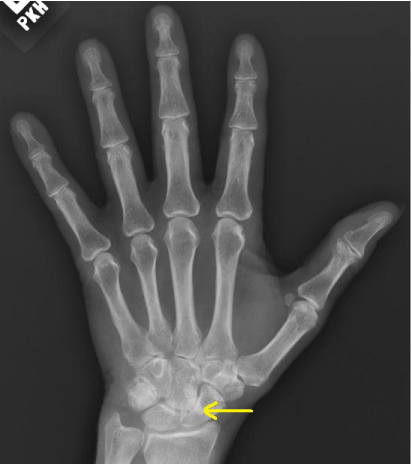
Roentgenography demonstrated a calcified mass overlying the scaphocapitate junction and
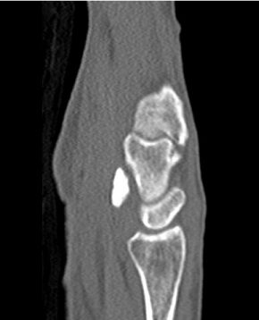
extending proximally (Figure 1). Subsequent computed tomography (CT) revealed a volar, densely
calcified RSC mass measuring 1.5 x 0.9 x 0.4 cm (Figure 2). Electrodiagnostic studies demonstrated
evidence of bilateral median neuropathies at the wrist, worse on the left. The patient was counseled
on operative versus continued non-operative management, and elected to pursue surgical removal
of the mass along with carpal tunnel release on the left.
Intra-operatively, the transverse carpal ligament was exposed with a standard incision along
the thenar crease. Its proximal and distal fibers were incised, openly exposing the carpal tunnel.
Significant tenosynovitis of the flexor tendons was noted. Inflamed synovium was dissected around
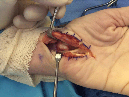
the tendons, with portions sent to pathology. Finally, the base of the carpal tunnel was visualized and the calcified mass was noted with a chalky, gouty appearance
(Figure 3). It was elevated and appeared to be originating from the
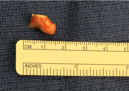
volar wrist. The calcified mass was excised from its origin (Figure 4)
and sent to pathology. Pathology established the calcareous material
to be non-polarizable.
Figure 1
Figure 1
Roentgenography demonstrating a calcified mass emanating from
the junction of the capitate and scaphoid.
Figure 2
Figure 3
Figure 4
Discussion
Amongst calcified masses in the wrist producing CTS, gout, pseudogout, tumoral calcinosis, and idiopathic calcification have been described [7-10]. Idiopathic calcified masses in the carpal tunnel are ascribed to patients with unilateral CTS, [6-10] acute CTS, [9] or systemic disease with propensity for ectopic calcification such as chronic renal failure, collagen vascular disease, and hypercalcemia [9,10]. The current study describes a patient with chronic bilateral CTS, exacerbated on the left by an idiopathic calcified cyst. Though our patient had a remote history of breast cancer status post mastectomy, there were no clinical findings to suggest active disease. The significant tenosynovitis of the FDS within the carpal tunnel likely contributed to the symptoms of CTS and may be present bilaterally. Whether there is a causative effect between the hyperplastic synovium of the FDS tendons and calcified cyst however, remains unknown. The course of the patient described in this study demonstrates that regardless of laterality, clinical suspicion for, and radiographic assessment of, mass-occupying lesions implicated in CTS should be undertaken where clinically appropriate.
References
- Ashworth NL. Carpal tunnel syndrome. BMJ Clin Evid. 2010; 2010: 1114.
- Palmer DH, Hanrahan LP. Social and economic costs of carpal tunnel surgery. Instr Course Lect. 1995; 44: 167-172.
- Cranford CS, Ho JY, Kalainov DM, Hartigan BJ. Carpal tunnel syndrome. J Am AcadOrthop Surg. 2007; 15: 537-548.
- Battaglia TC, Pannunzio ME, Chhabra AB. A carpometacarpal joint ganglion cyst causing median neuropathy. Am J Orthop. 2006; 35: 186- 188.
- Plate AM, Lee SJ, Steiner G, Posner MA. Tumor like lesions and benign tumors of the hand and wrist. J Am Acad Orthop Surg. 2003; 11: 129-141.
- Nakamichi K, Tachibana S. Unilateral carpal tunnel syndrome and spaceoccupying lesions. J Hand Surg Br. 1993; 18: 748-749.
- Takada T, Fujioka H, Mizuno K. Carpal tunnel syndrome caused by an idiopathic calcified mass. Arch Orthop Trauma Surg. 2000; 120: 226-227.
- Sensui K, Saitoh S, Kametani K, Makino K, Ohira M, Kimura T, et al. Property analysis of ectopic calcification in the carpal tunnel identification of apatite crystals: a case report. Arch Orthop Trauma Surg. 2003; 123: 442-445.
- Verfaillie S, De Smet L, Leemans A, Van Damme B, Fabry G. Acute carpal tunnel syndrome caused by hydroxyapatite crystals: a case report. J Hand Surg Am. 1996; 21: 360-362.
- Cofan F, Garcia S, Combalia A, Segur JM, Oppenheimer F. Carpal tunnel syndrome secondary to uraemictumoral calcinosis. Rheumatology (Oxford). 2002; 41: 701-703.


