Case Report
Unexpected Foreign Body in Neck: A Case Report
Ramdas Balakrishna, Anubhav Jannu*, Sudarshan, Veena GC and Bhuvaneshwari
Department of Oral and Maxillofacial Surgery, KLE Dental College and Hospital & Research Centre, India
*Corresponding author: Anubhav Jannu, Department of Oral and Maxillofacial Surgery, KLE Dental College and Hospital & Research Centre, No 20, Yeshwanthpur Suburb, Bangalore-560022, Karnataka, India
Published: 07 Mar, 2017
Cite this article as: Balakrishna R, Jannu A, Sudarshan,
Veena GC, Bhuvaneshwari.
Unexpected Foreign Body in Neck: A
Case Report. Ann Clin Case Rep. 2017;
2: 1290.
Abstract
Foreign bodies may be deposited, ingested or inserted in the head and neck region by a traumatic or iatrogenic injury. The penetrating foreign body in the neck has a specific apprehension because of the constellations of vital structures in the neck. We report a 30 year old male patient who presented with a mass in the neck. Radiographic examination revealed a sharp No.11 surgical blade in the left side of the neck. He was unaware of the foreign body in his neck and was asymptomatic. The blade was surgically removed followed by primary closure under general anesthesia. The patient recovered well without any complications.
Keywords: Orthopantamogram; Suprahyoid; Hemodynamically; Foreign bodies
Introduction
Deposited foreign bodies are not so common problem among any age group. In the head and neck region these foreign bodies are interesting and at times challenging to locate and remove [1- 3]. They may be deposited by a traumatic or iatrogenic injury. The case described here is a report of a deposited No.11 surgical blade into the neck, which the patient was unaware off [4]. This case highlights the possibility that relatively big sharp blade can also penetrate the neck and present as a foreign body in the neck.
Case Presentation
A 30-year-old male patient was referred to our institution with a firm fibrous mass in
the left side of the neck. He was asymptomatic, but gives a history of swelling since 5 years. An
orthopantamogram revealed a foreign body (No.11 surgical blade) with prominent margins inferior
to the base of the mandible and superior to the suprahyoid muscle in the left side of the neck (Figure 1), being completely embedded in the soft tissue. No evident scars were present on the skin and
no fistula was found with the deeper tissues. The patient gives a history of extraction of lower left
first molar tooth, which was done smooth and non-surgically without the use of surgical blade. The
patient also gives a history of trauma 5 years ago with laceration, which was sutured in a private
hospital under local anesthesia. Hence, the patient is unaware of the surgical blade (No.11) in the
left side of his neck.
Physical examination revealed a firm fibrotic mass measuring 2 cms x 2 cms in the left side of his
neck. There was no tenderness or pain in and around that region. The rest of the Head and Neck did
not reveal any scars or abnormalities. The blood samples revealed everything normal. The patient
was hemodynamically stable.
The patient was hospitalized after initial examination, followed by taking orthopantamogram
and an informed consent. In the operation theatre under general anesthesia, the mass was explored
by a submandibular incision perpendicular to the long axis of the foreign body on the neck.
After sub-platysmal dissection, the surgical blade was identified (Figure 2). All the fibrotic bands
around the blade were dethatched. It had not damaged any vital structures. The surgical blade was
completely exposed followed by the identification of the broken tip initially and later removal of the
entire blade (Figure 3). A good primary closure was achieved. Intravenous antibiotics, analgesics
and prophylaxis of tetanus were administered in appropriate doses. Follow-up after the 3rd, 7th and
15th day revealed complete healing of the wound.
Figure 1
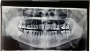
Figure 1
Orthopantamogram showing surgical blade in the left side of the
neck.
Figure 2
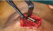
Figure 2
Intra-operative picture showing surgical blade in the left side of
the neck.
Figure 3
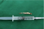
Figure 3
Two pieces of the surgical blade recovered after surgery.
Discussion
Since ages, deposited or impacted foreign bodies have been a less amusement to the clinicians
as well as to the population. Sometimes it may turn out uneventfully, otherwise it endangers the
life of the patient depending on the type, size and location of the foreign body [2,5]. Deposition or
impaction of foreign bodies is a very scary situation for the patient as well as their close ones. They are usually secondary to a gunshot or stab wound [6,7]. There have
been reports of impacted chopsticks [8], pen [6] and wooden piece
[2]. However, retention of a surgical blade (No.11) is less reported.
The diagnosis of penetrating neck trauma with an associated foreign
body in situ is generally quite obvious from history or clinical
examination. However identifying a foreign body can be very
challenging at times, especially in cases where the impacted body is
very thin or radiologically not very clear [6,9]. Precise localization of
the foreign object is essential for complication free removal [6]. The
current mortality rate for penetrating neck injury is 3-6%. The usual
complications of penetrating neck injuries is less than 10% to as high
as 20% and a mortality rate of as high as 20% is also reported [2,5,10].
The present case is interesting because of the mode of entry of the
surgical blade in the neck, which the patient has been unaware off. It
is also interesting in the way it travels under the subcutaneous tissue
of the neck without creating any complications. It was detected with an orthopantamogram. It traveled sub platysmally and was stuck 5
mm below the base of the mandible on the left side of the neck. To
prevent any complications intra-operatively or post- operatively, the
wound should be explored by horizontal incision in the skin crease
with proper wound debridement.
Thorough knowledge of the anatomy of the neck, physical
examination and current recommendations for diagnostics and
therapeutic interventions are necessary for the appropriate removal
of the foreign bodies in the neck region [2].
Conclusion
Foreign bodies in the neck are an uncommon but potentially life threatening and crisis condition. Pre-operative imaging is very important in deciding upon the surgical approach for the retrieval of impacted foreign bodies. A thorough knowledge of the anatomy, and various management protocols with a changing technique compel the surgeon to perform a close evaluation of the patient. Each maneuver should be directed to reduce the rate of morbidity and mortality by means of timely intervention. In cases of surgical blade as foreign bodies, early exploration and surgical removal reduces the chances of fibrosis, infection and damage to vital structures, resulting in a favorable outcome.
References
- Hunter TB, Taljanovic MS. Foreign bodies. Radiographics. 2003; 23: 731- 757.
- Rakesh KS, Sangita B, Prahlad K. Managing a wooden foreign body in the neck. J Emerg Trauma Shock. 2009; 2: 191-195.
- Dua R, Morgan N, Kichenaradjou. Foreign bodies. 2011; 40: 1149.
- Ozturk k, Turhal G, Gode S, Atilla Yavuzer. Migration of a swallowed blunt foreign body to the neck. Case reports in Otolaryngology. 2014; ID 646785: 2.
- Kendall JL, Anglin D, Demetriades D. Penetrating Neck Trauma. Emerg Med Clin North Am.1998; 18: 85-105.
- Latha PR, Sherry P, Sreekumar KP, Subramaniya Iyer. A pen in the neck : An unusual foreign body and an unusual path of entry. Indian J Dent Res. 2014; 25: 111-114.
- Wakisaka H, Takahashi H, Ugumori T, Motoyoshi K, Daiki Takagi A case of wooden foreign body penetrating the oral cavity and reaching the posterior neck. Inj Extra. 2010; 41: 92-96.
- Park SH, Cho KH, Shin YS, Kim SH, Ahn YH, Cho KG, et al. Penetrating Craniofacial Injuries in Children with wooden and metal chopsticks. Pediatr Neurosurg. 2006; 42: 138-146.
- Hersman G, Barker P, Bowley DM, Boffard KD. The management of penetrating neck injuries. Int Surg. 2001; 86: 82-89.
- Nason RW, Assuras GN, Gray PR, Lipschitz J, Burns CM. Penetrating neck injuries: Analysis of experiences from a Canadian trauma center. Can J Surg.2001; 44: 122-126.
- Gracias VH, Reilly PM, Philpott J, Klein WP, Lee SY, Singer M, et al. Computed tomography in the evaluation of penetrating neck trauma, Arch Surg. 2001; 136: 1231-1235.