Case Report
Spigelian Hernia: A Rare Case Report
Wael Zaki and Awad Ali M Alawad*
Department of Surgery, Prince Sultan Armed Forces Hospital, Saudi Arabia
*Corresponding author: Awad Ali M. Alawad, Department of Surgery, Prince Sultan Armed Forces Hospital, Medina, Saudi Arabia
Published: 06 Mar, 2017
Cite this article as: Zaki W, Alawad AAM. Spigelian Hernia:
A Rare Case Report. Ann Clin Case
Rep. 2017; 2: 1288.
Abstract
Spigelian hernias are rare abdominal wall defects that occur at the semilunar line lateral to the rectus abdominis muscle. They are located between the muscular layers of the abdominal wall and can be easily overlooked because of abdominal obesity. Generally, they are difficult to diagnose because of their location and vague symptoms. The diagnosis has been considerably aided by the introduction of ultrasonography and Computed Tomography. Once the diagnosis is made operative management is indicated due to risk of incarceration. We report a 32 years old female patient from who presented with right upper abdominal pain associated with a swelling below the right subcostal margin. A diagnosis of Spigelian hernia and gallbladder stones was made. The patient underwent laparoscopic mesh repair and cholecystectomy. Her recovery was uneventful.
Keywords: Spigelian hernia; Lateral ventral hernias; Laparoscopic mesh repair
Introduction
Spigelian hernia is named after Adrian Van der Spighel who described semilunar like
(lineaspigeli) in 1645. The hernia was first described Klinkosch in 1764 [1]. Spigelian hernia is a
rare abdominal hernia, occurring through the spigelianaponeurosis, it carries a significant risk
of incarceration and strangulation. Most spigelian hernias occur below the level of the umbilicus
close to the level of the arcuate line (inferior margin of posterior leaflet of rectus sheath within the
abdomen), though they have being reported to occur above the level of the umbilicus [2].
Diagnosis of Spigelian hernia requires a high degree of suspicion, with the most common
finding on clinical examination being a lump at the semilunar line. Radiological tests are useful
in confirming the diagnosis. Once diagnosed, Spigelian hernias require operative repair. Elective
repair of uncomplicated Spigelian hernias can be performed both laparoscopically and by an open
technique, with the former reported to be associated with a lower morbidity and shorter hospital
stay [3]. We present a case of spigelian hernia in a female patient and its management and discuss
about the various investigations and the treatment modalities available for its repair, with literature
review.
Case Presentation
A 32 years old female patient presented to the outpatient department with history of intermittent
right upper quadrant pain for 8 years. She underwent laparoscopic sleeve gastrectomy 2 years ago.
On examination, she had a weight of 89 kg with a BMI of 33 kg/m2. Physical examination revealed
a firm lump of about 6 x 5 cm was found on the right upper abdomen at the margin of the right
semilunar line. The lump would increase on coughing and decrease in lying position.
On investigation, haemogram, liver function tests, blood urea and creatinine were normal.
Ultrasonography showed multiple gall stones and right hypochondrium anterior abdominal wall
defect (Figure 1). CT scan showed the defect of a Spigelian hernia about 3 cm in diameter, containing omentum (Figure 2).
After adequate preparation she was planned for laparoscopic cholecystectomy and hernia repair.
Intraoperatively, right sided SH was detected. There was also hemangioma on the lower part of the
right lobe. Laparoscopic cholecystectomy was done and the hernia was managed laparoscopically.
Omentum adherent to the defect was reduced. The defect was seen as a large opening in the
peritoneum, along the lateral margin of rectus abdomens muscle on the right side (Figure 3). After dissection of the adhesions with the help of Harmonic scalpel, a prosthetic composite mesh (10 x 10
cm) was introduced into the peritoneal cavity and was fixed with the help of tacks to cover the defect
(Figure 4). A full laparoscopic exploration of the abdomen was completed without finding other
defects. Postoperative recovery was uneventful and the patient was discharged on postoperative
day 2.
Figure 1
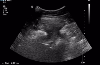
Figure 1
Right hypochondrium anterior abdominal wall defect averaging 2.6
cm with omental content.
Figure 2
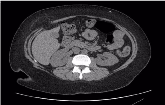
Figure 2
CT scan showing the defect of a Spigelian hernia about 3 cm in
diameter, containing omentum.
Figure 3
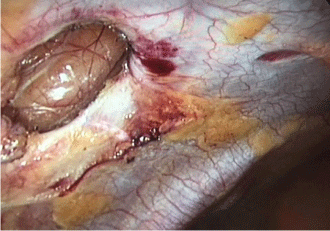
Figure 3
Laparoscopic image showing the defect of a Spigelian hernia and
the previous port.
Figure 4
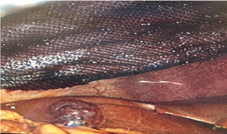
Figure 4
Laparoscopic image showing the defect of a Spigelian hernia and
the previous port.
Discussion
Since ages, deposited or impacted foreign bodies have been a less amusement to the clinicians
as well as to the population. Sometimes it may turn out uneventfully, otherwise it endangers the
life of the patient depending on the type, size and location of the foreign body [2,5]. Deposition or
impaction of foreign bodies is a very scary situation for the patient as well as their close ones. They are usually secondary to a gunshot or stab wound [6,7]. There have
been reports of impacted chopsticks [8], pen [6] and wooden piece
[2]. However, retention of a surgical blade (No.11) is less reported.
The diagnosis of penetrating neck trauma with an associated foreign
body in situ is generally quite obvious from history or clinical
examination. However identifying a foreign body can be very
challenging at times, especially in cases where the impacted body is
very thin or radiologically not very clear [6,9]. Precise localization of
the foreign object is essential for complication free removal [6]. The
current mortality rate for penetrating neck injury is 3-6%. The usual
complications of penetrating neck injuries is less than 10% to as high
as 20% and a mortality rate of as high as 20% is also reported [2,5,10].
The present case is interesting because of the mode of entry of the
surgical blade in the neck, which the patient has been unaware off. It
is also interesting in the way it travels under the subcutaneous tissue
of the neck without creating any complications. It was detected with an orthopantamogram. It traveled sub platysmally and was stuck 5
mm below the base of the mandible on the left side of the neck. To
prevent any complications intra-operatively or post- operatively, the
wound should be explored by horizontal incision in the skin crease
with proper wound debridement.
Thorough knowledge of the anatomy of the neck, physical
examination and current recommendations for diagnostics and
therapeutic interventions are necessary for the appropriate removal
of the foreign bodies in the neck region [2].
Conclusion
Foreign bodies in the neck are an uncommon but potentially life threatening and crisis condition. Pre-operative imaging is very important in deciding upon the surgical approach for the retrieval of impacted foreign bodies. A thorough knowledge of the anatomy, and various management protocols with a changing technique compel the surgeon to perform a close evaluation of the patient. Each maneuver should be directed to reduce the rate of morbidity and mortality by means of timely intervention. In cases of surgical blade as foreign bodies, early exploration and surgical removal reduces the chances of fibrosis, infection and damage to vital structures, resulting in a favorable outcome.
References
- Hunter TB, Taljanovic MS. Foreign bodies. Radiographics. 2003; 23: 731- 757.
- Rakesh KS, Sangita B, Prahlad K. Managing a wooden foreign body in the neck. J Emerg Trauma Shock. 2009; 2: 191-195.
- Dua R, Morgan N, Kichenaradjou. Foreign bodies. 2011; 40: 1149.
- Ozturk k, Turhal G, Gode S, Atilla Yavuzer. Migration of a swallowed blunt foreign body to the neck. Case reports in Otolaryngology. 2014; ID 646785: 2.
- Kendall JL, Anglin D, Demetriades D. Penetrating Neck Trauma. Emerg Med Clin North Am.1998; 18: 85-105.
- Latha PR, Sherry P, Sreekumar KP, Subramaniya Iyer. A pen in the neck : An unusual foreign body and an unusual path of entry. Indian J Dent Res. 2014; 25: 111-114.
- Wakisaka H, Takahashi H, Ugumori T, Motoyoshi K, Daiki Takagi A case of wooden foreign body penetrating the oral cavity and reaching the posterior neck. Inj Extra. 2010; 41: 92-96.
- Park SH, Cho KH, Shin YS, Kim SH, Ahn YH, Cho KG, et al. Penetrating Craniofacial Injuries in Children with wooden and metal chopsticks. Pediatr Neurosurg. 2006; 42: 138-146.
- Hersman G, Barker P, Bowley DM, Boffard KD. The management of penetrating neck injuries. Int Surg. 2001; 86: 82-89.
- Nason RW, Assuras GN, Gray PR, Lipschitz J, Burns CM. Penetrating neck injuries: Analysis of experiences from a Canadian trauma center. Can J Surg.2001; 44: 122-126.
- Gracias VH, Reilly PM, Philpott J, Klein WP, Lee SY, Singer M, et al. Computed tomography in the evaluation of penetrating neck trauma, Arch Surg. 2001; 136: 1231-1235.