Case Report
Visual Loss in Raised Intracranial Pressure Associated with Severe Vitamin A Deficiency
Olaoluwakitan A Osunkunle1, Robert MJ Purbrick1,2 and Susan M Downes1,3*
1Oxford Eye Hospital, Oxford University Hospitals NHS Foundation Trust, UK
2Sussex Eye Hospital, Brighton and Sussex University Hospitals NHS Trust, UK
3Nuffield Department of Ophthalmology, University of Oxford, UK
*Corresponding author: Susan M Downes, Oxford Eye Hospital, Oxford University Hospitals NHS Foundation Trust, West Wing, John Radcliffe Hospital, Headley Way, Oxford OX3 9DU, UK
Published: 08 Feb, 2017
Cite this article as: Osunkunle OA, Purbrick RMJ, Downes
SM. Visual Loss in Raised Intracranial
Pressure Associated with Severe
Vitamin A Deficiency. Ann Clin Case
Rep. 2017; 2: 1261.
Abstract
Idiopathic Intracranial Hypertension (IIH) is a disorder characterized by raised intracranial
pressure without evidence of another cause such as infection, space occupying lesion or vascular
abnormality. The incidence of IIH seems to be increasing amongst adolescent children, but the
pathogenesis of the disease still remains unclear.
We report a case of visual loss with raised intracranial pressure, and retinal dysfunction associated
with unrecordable levels of vitamin A in a child. A twelve year old boy presented with reduced
vision; on examination there was bilateral papilloedema. Investigations showed raised intra-cranial
pressure and rod-cone dysfunction on electrophysiology but normal appearances on the CT and
MRI head.
This pathologic pattern was attributed to idiopathic intracranial hypertension associated with
severe Vitamin A deficiency. This case highlights the importance of considering malnutrition, when
confronted with the dual presentation of raised intracranial pressure and retinal dysfunction.
Introduction
This case report describes an association between hypovitaminosis A and raised intracranial
pressure in a 12 year old boy. Vitamin A belongs to a family of lipid soluble compounds known as
retinoic acids. It exists in two main forms: provitamin A carotenoids such as beta-carotene, which
are found in plants and preformed vitamin A, retinols, found in animal sources of food. Animal
sources containing relatively high levels of retinols include: liver, kidney, egg yolk and butter.
The classical ocular presentation of vitamin A deficiency is known as xerophthalmia. The initial
finding is that of dryness of the conjunctiva and cornea because of a reduction in lacrimal gland
secretions. Bitot's spots, which are abnormal squamous cell proliferations with keratinization of the
conjunctiva, may then occur. The eye may progress to corneal xerosis and keratomalacia. There is
also visual pigment dysfunction. This may result in nyctalopia (night blindness) and retinopathy.
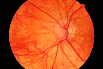
Figure 1
Results and Discussion
A 12 year old male presented with a two week history of painless loss of vision. There was no relevant family or previous history apart from a diagnosis of a mild autistic spectrum disorder. His diet comprised white bread, crisps, chocolate, biscuits and French fries. His daily energy intake was about 2500 kcal/day. His weight was 61.3 kg (99.6th percentile). Vision was decreased to 6/12 right and hand movements left. He had a left relative afferent pupillary defect; bilateral reduced colour vision with no Ishihara plates seen by the left eye. Moderate bilateral disc swelling (Figure 1) was present, with no other ocular abnormalities. CT and MRI scans of his head showed no mass lesions or venous sinus thrombosis. A lumbar puncture showed a raised opening pressure of 45 cm H20; analysis of cerebrospinal fluid analysis showed a glucose of 3.4 mmol/L with a mildly elevated protein of 442 mg/L and no antibodies to myelin oligodendrocyte glycoprotein or aquaporin 4. Intravenous acetazolamide was started for IIH. Blood screen was normal apart from the vitamin A, which was undetectable at <0.5 μmol/L (range: 0.92 – 1.71). He started paediatric seravit, 25 gms daily orally (Vitamin A 10 000IU). Electrophysiology was consistent with rod-cone dysfunction. Fields were constricted bilaterally with a left centrocecal scotoma. By four weeks, his vitamin A levels had normalised at 1.26 μmol/L and visual acuities had improved to 6/6 right and counting fingers left. Repeat electrophysiology at three months showed bilateral significant improvement.
Conclusion
IIH is more commonly described in association with excess rather than a deficiency of vitamin A [1,2]. Other causes include drugs, hormonal and metabolic imbalances and toxins [2]. None of these was relevant, although our patient was on the 99.6th centile for weight. His diet was essentially vegan with no fresh vegetables, and his rigid choice of food was attributed to his mild autism disorder. He did not have dry eye symptoms or ocular signs of vitamin A deficiency (VAD). VAD associated with IIH has been described in a paediatric population before [3]. VAD is common worldwide, but it is rare in industrialised countries and when found in the developed world may be associated with patients with mental health disorders [4]. IIH and VAD secondary to a restricted diet in autistic patients has been described [3] but unlike this case, they did not have retinal dysfunction. In VAD rat and calves, electron microscopy has revealed fibrotic changes in the arachnoid villi [5]. An inability to absorb CSF properly could be an explanation for raised intracranial pressure. This case highlights the importance of asking about diet in a child who has autism presenting with IIH to check for retinal dysfunction, and to check vitamin A levels. It is also important to realize that corneal signs may not be present in VAD.
References
- Rangwala LM, Liu GT. Pediatric idiopathic intracranial hypertension. Surv Ophthalmol. 2007; 52: 597-617.
- Wall M. Idiopathic intracranial hypertension. Neurol Clin. 2010; 28: 593- 617.
- Dotan G, Goldstein M, Stolovitch C, Kesler A. Pediatric pseudotumor cerebri associated with low serum levels of vitamin A. J Child Neurol. 2013; 28: 1370-1377.
- Lewis CD, Traboulsi EI, Rothner AD, Jeng BH. Xerophthalmia and Intracranial Hypertension in an Autistic Child with Vitamin A Deficiency. J Pediatr Ophthalmol Strabismus. 2010; 48: e1-3.
- Hayes KC, McCombs HL, Faherty TP. The fine structure of vitamin A deficiency II. Arachnoid granulations and CSF pressure. Brain. 1971; 94: 213-224.

