Case Report
Skull Base Inverted Papilloma: A Case Report
Shalaka Sharma1*, Divij Sonkhya2 and Nishi Sonkhya3
1Department of Otolaryngology, SMS Medical College and Hospital, India
2Department of Otolaryngology, Sonkhya Hospital, India
3Department of Otolaryngology, SMS Medical College and Hospital, India
*Corresponding author: Shalaka Sharma, Department of Otolaryngology, SMS Medical College and Hospital, India
Published: 21 Dec, 2016
Cite this article as: Sharma S, Sonkhya D, Sonkhya N.
Skull Base Inverted Papilloma: A Case
Report. Ann Clin Case Rep. 2016; 1:
1219.
Abstract
Inverted papilloma is an uncommon primary nasal. Despite its benign nature, this tumor represents three typical characteristics: a high propensity of recurrence, local aggressiveness and association with malignancy. Inverted papilloma can reduce the patient’s quality of life due to compromised nasal function, extension to orbit and brain even in absence of malignancy. We are presenting a case of 66 year old male patient with bilateral inverted papilloma with intracranial extensions after multiple recurrences operated 10 times over a period of 28 years, with extensive resection of disease every time but without any evidence of malignancy till date still showing fast and aggressive recurrences.
Introduction
Inverted papilloma is a benign epithelial growth extending into the underlying stroma of the nasal cavity and paranasal sinus. These tumors arise from the Schneiderian respiratory membrane that lines the nasal cavity and paranasal sinuses [1]. Schneiderian papillomas can be classified in into 3 types: exophytic (everted or fungiform), inverted, and oncocytic (columnar or cylindrical) [2]. In 1854 Ward first reported this type of tumor in nasal cavity [3]. Ringertz et al. [4] in 1938 was the first to identify endophytic growth patterns of IPs with its characteristic tendency to invert into underlying stroma and called it “inverted papilloma”. Hyams [2] divided papillomas of sinonasal tract into three histological categories due to their pattern of growth: (a) Fungi form (everted) papilloma, (b) Oncocytic schneiderian papillomas, (c) Inverted papillomas. Inverted papilloma (IP) is an uncommon lesion that accounts for 0.5–4% of all primary nasal tumors [5]. It affects all ages, most commonly males [6] in the fifth to the seventh decades of life [7]. The most frequent sites are the lateral nasal wall near the middle turbinate or ethmoid recesses and the maxillary sinuses. The nasal septum, frontal and sphenoid sinuses are rarely affected. Common sites of intracranial spread include the cribriform plate, fovea ethmoidalis, and orbits [8]. The tumor is well known for its invasiveness, tendency to recur and association with malignancy [9].
Case Presentation
AA 66 year old Hindu male patient presented to department of ENT of SMS hospital with few
months history of bilateral nasal fullness, purulent nasal discharge, posterior nasal drip, proptosis of
left eye and headache. No history of tobacco smoking, alcohol intake or allergy was present.
Over past 28 years patient was operated 10 times for bilateral nasal mass and different
approaches were used at different centers by experienced surgeons. Patient underwent Transnasal
resection of tumor twice (year1989, 1991), Moore’s lateral rhinotomy excision of tumor (year1993,
1995), Midfacial degloving approach (year1999), Endoscopic excision of tumor (year2001, 2003),
Bilateral lateral rhinotomy with craniofacial resection (year2005), Endoscopic excision with frontal
sinustomy by external approach (year2009).
Histopathological diagnosis of the first operation was nonspecific inflammation of the nasal
mucosa, while the pathological diagnosis of rest of the surgeries was Inverted Papilloma. Patient was
also advised radiotherapy but he refused for it.
Clinical examination of the patient revealed left eye proptosis with diplopia of left eye, left side
frontal bossing, and normal vision. There was no cranial nerve palsy no cervical lymphadenopathy.
On performing Anterior Rhinoscopy we found single nasal cavity with absence of nasal septum.
Minimal lesion was present along lateral nasal wall. Nasal endoscopy showed single nasal cavity
with absence of nasal septum, middle turbinate, inferior turbinate. Papilloma like mass along lateral nasal wall and a cystic mass seen hanging from roof of nasal cavity.
Since recurrent Inverted Papilloma was suspected patient was
elected for CT and MRI of head and neck areas which showed
heterogeneous opacification of mass in right maxillary sinus, with
minimal lesion in left maxillary sinus and nasal cavity. Nasal septal
perforation due to prior endonasal resections was also observed
(Figure 3). Cystic mass occupying frontoethmoid region with
intracranial extension (Figure 1 and 2).
Under general anesthesia patient underwent endoscopic
excision of tumor from lateral nasal wall and maxillary sinus along
with aspiration of fluid from the cyst. Histopathological study of
the resected specimen as well as cytology of the fluid confirmed the
diagnosis of Inverted Papilloma without evidence of malignancy.
Further HPV studies of the patient came negative.
Figure 1
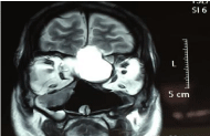
Figure 1
Coronal section MRI of nose and pns showing cystic mass
occupying frontoethmoid region with intracranial extension (arrow).
Figure 2
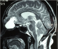
Figure 2
MRI saggital cuts of nose and paranasal sinus showing cystic
mass occupying frontoethmoid region with intracranial extension.
Figure 3
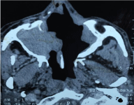
Figure 3
Contrast enhanced CT axial sections showing single nasal cavity
with minimal lesion along lateral nasal wall and maxilla.
Discussion
Inverted papilloma can be defined as a group of benign neoplasm
arising from the sinonasal (Schneiderian) mucosa and is composed
of squamous or columnar epithelial proliferation with associated
mucous cells. Schneiderian papillomas represent <5% of all sinonasal
tract tumors. Inverted papillomas occur along the lateral nasal wall
(middle turbinate or ethmoid recesses), with secondary extension into
the paranasal sinuses. They may originate in paranasal sinus with/
without involving nasal cavity. Typically, the schneiderian papillomas
are unilateral; bilateral papillomas may also occur. In our case it was bilateral inverted papilloma since beginning. Symptoms vary
according to the site of occurrence and include airway obstruction,
epistaxis and a symptomatic mass or pain.
The etiology of inverted papilloma is still unclear. There have
been many causes suggested such as allergy, chronic sinusitis, viral
infections, and inflammation, HPV [10].
Significant association has been identified between the presence
of human papilloma virus DNA in inverted papilloma and recurrence
after surgical resection [11]. HPV 16 and 18 were found to be related to
the malignant transformation of Inverted Papilloma [12]. In addition
to HPV, studies have showed that p21 and p53, probably coupled
epidermal growth factor receptor (EGFR; ErbB-1), transforming
growth factor alpha (TGF-α), Topoisomerase II- alpha are predictive
of malignant transformation [13]. The observation that Inverted
Papilloma tend to recur after incomplete surgical removal supports
investigation suggested that Inverted Papilloma is a true neoplasm
arising from single progenitor cell and the recurrence represents
growth of the residual clone [14]. In our study we found that the
patient was HPV negative and molecular studies could not be done
because of unavailability of such studies at our centre but in future if
such studies are available then we will get them done.
Differential diagnosis of Inverted Papilloma includes antrochoanal
polyp, nasal cavity squamous polyp, allergic fungal sinusitis, fibrous
dysplasia, giant cell granuloma, juvenile angiofibroma, nasal glioma,
meningoencephalocele, mucocele, mucus retention cyst, Thornwaldt’s
cyst, grossly enlarged adenoids, SCC, lymphoma, adenocarcinoma,
esthesioneuroblastoma. Sinonasal inflammatory polyps are clinically
similar but histopathologically epithelial alterations are seen in
inverted papillomas and not in the inflammatory polyps. Unilateral
tumor localization involving the lateral nasal wall and the middle
meatus is a diagnostic clue to Inverted Papilloma.
CT and MRI are techniques of choice for pretreatment staging
in IP. Due to nature of tumor histology occurring in paranasal
sinuses, CT offers superior bony definitions and MRI gives superior
soft tissue delineation [15]. Unilateral opacification of the paranasal
sinuses is typical CT finding of inverted papilloma. Bony changes on
CT imaging of inverted papilloma are useful for predicting tumor
origin and recurrence sites. Focal hyperostosis, bony struts or osteitis
detected on preoperative CT can predict with high degree of accuracy
the site of origin [16]. Erosion, remodeling and widening of the natural orifices of the sinuses on a CT scan are useful signs indicating IP.
MRI is superior to CT scan in distinguishing papillomas from
inflammation. With gadolinium enhancement, MRI demonstrates
perineural invasion and dural or intracranial involvement very well.
MRI is useful for planning an appropriate surgical approach, and
for selecting cases that can be managed by endoscopic approaches,
resulting in lower rates of tumorrecurrence and morbidity [17].
Dynamic MR imaging can differentiate accurately recurrent IP
from postoperative changes. On T1- weighted images, sinonasal
papillomas appear slightly hyperintense to muscle; on T2- weighted
images, sinonasal papillomas have intermediate intensity.
Intracranial extension and dural penetration is rare and often
associated with recurrent disease [18]. Tumor may spread by direct
invasion of bone and cartilage to involve related structures, but the
walls of the nasal cavity and paranasal sinuses also contain numerous
foraminae and fissures transmitting important neurovascular
bundles.
The primary and preferred treatment of inverted papilloma is
surgery. Precise determination of sites tumor origin and attachment
during the operation, strict application of selection criteria, proper
preoperative evaluations, intra-operative determination of extent
and attachment of the tumor, close endoscopic follow up, expert
application of endoscopic techniques, meticulous use of subperiosteal
dissection in the involved areas, wide removal of the tumor origin
along the subperiosteal plane as well as drilling the underlying bone
[19], complete removal of all the diseased mucosa with creation of
wide cavities, and long term regular follow up evaluation are the
key elements to the successful treatment. Limited involvement of
the skull base can be successfully achieved by endoscopic excision.
Endoscopic resection is associated with shorter hospital stay, shorter
operative time and lesser morbidity. External approach has been gold
standard for sinonasal tumor removal [20], but is associated with
several side effects, including facial scars, intracranial and extracranial
complications, long hospital stay. The external surgical approach is
adequate for tumors extending to the brain, orbit and maxillary sinus.
“Recurrence” actually represents residual disease in most cases.
The magnitude of the recurrence is directly proportional to the
completeness of removal with the best results obtained by techniques
that afford the best operative exposure.
There is also association of malignancy with inverted papilloma.
Malignancy was found to be associated with bilateral inverted
papilloma, histologic multicentricity, a predominance of mature
squamous epithelium, severe hyperkeratosis, absence of inflammatory
polyps among the papillomas. In our case it was bilateral inverted
papilloma since beginning with presence of focal squamous epithelial
cells in HPR with 10 times recurrence over 28 years but still there
were no signs of malignancy.
Conclusion
This case report presents Inverted papilloma with intracranial extension after multiple recurrences. Here the Inverted papilloma was bilateral which is less common and a risk factor for malignancy but still no malignancy reported till date. High rate of recurrence, local aggressiveness and association of malignancy makes it necessary to perform radical surgery for this benign tumor. Aggressive treatment of intranasal inverted papilloma is the most important factor in preventing intracranial presentation. Because of the malignant potential of the disease long term follow up of the disease is also mandatory. HPV studies came negative in our case, but due consideration must be given to such studies i.e. HPV and molecular studies so that a better understanding of alterations in epithelial cell proliferation and cell cycle regulation in inverted papilloma may lead to adjuvant medical therapies to decrease recurrence rates and improve treatment.
References
- Figuerola E, Liern M, Palomero JM. Nasosinusal inverted papilloma. Acta Otorrinolaringologicaespanola. 1990; 41: 35-38.
- Hyams VJ. Papillomas of the nasal cavity and paranasal sinuses. A clinicopathological study of 315 cases. Ann Otol Rhinol Laryngol. 1971; 81: 192-206.
- Ward N. A mirror of the practice of medicine and surgery in the hospitals of London. The Lancet. 1854; 2: 480-482.
- Ringertz N. Pathology of malignant tumors arising in the nasal and paranasal cavities and maxilla. Acta Otolaryngol. 1938; 27: 31-42.
- Vrabec DP. The inverted Schneiderian papilloma: A 25-year study. Laryngoscope. 1994; 104: 582–605.
- Lesperance MM, Esclamado RM. Squamous cell carcinoma arising in inverted papilloma. Laryngoscope. 1995; 105: 178-183.
- Segal K, Atar E, Mor C, Har-El G, Sidi J. Inverting papilloma of the nose and paranasal sinuses. Laryngoscope. 1986; 96: 394-398.
- Visvanathan V, Wallace H, Chumas P, Makura ZG. An unusual presentation of inverted papilloma: case report and literature review. J Laryngol Otol. 2009; 124: 101-104.
- Mirza S, Bradley PJ, Acharya A, Stacey M, Jones NS. Sinonasal inverted papillomas: recurrence, synchronous and metachronous malignancy. J J Laryngol Otol. 2007; 121: 857-864.
- Lawson W, Schlecht NF, Brandwein M, Gensler. The role of the human papilloma virus in the pathogenesis of Schneiderian inverted papillomas: an analytic overview of the evidence. Head Neck Pathol. 2008; 2: 49-59.
- Beck JC, Mc Clatchey KD, Lesperance MM, Esclamado RM, Carey TE, Bradford CR. Presence of human papilloma virus predicts recurrence of inverted papilloma. Otolaryngol Head Neck Surg. 1995; 113: 49-55.
- Zhong Z, Yan A, Jiang F, Wei H, Jiang X. The study on the relationship between human papilloma virus infection and pathogenesis of nasal inverted papilloma and its malignant transformation. Lin Chuang Er Bi Yan Hou Tou Jing Wai Ke Za Zhi. 2010; 24: 209-211.
- Altavilla G, Staffieri A, Busatto G, Canesso A, Giacomelli L, Marioni G. Expression of p53, p16INK4A,pRb, p21WAF1/CIP1, p27KIP1, cyclin D1, Ki-67 and HPV DNA in sinonasal endophytic Schneiderian (inverted) papilloma. Acta Otolaryngol. 2009; 129: 1242-1249.
- Califano J, Koch W, Sidransky D, Westra WH. Inverted sinonasal papilloma: a molecular genetic appraisal of its putative status as a precursor to squamous cell carcinoma. Am J Pathol. 2000; 156: 333-337.
- Loevner LA, Sonners AI. Imaging of neoplasms of the paranasal sinuses. Neuroimaging Clin N Am. 2004; 14: 625-646.
- Lee DK, Chung SK, Dhong HJ, Kim HY, Kim HJ, Bok KH. Focal hyperostosis on CT of sinonasal inverted papilloma as a predictor of tumor origin. Am J Neuroradiol. 2007; 28: 618-621.
- Roobottom CA, Jewell FM, Kabala J. Primary and recurrent inverting papilloma: appearances magnetic resonance imaging. Clin Radiol. 1995; 50: 472-475.
- Vural E, Suen JY, Hanna E. Intracranial extension of inverted papilloma: an unusual and potentially fatal complication. Head Neck. 1999; 21: 703- 706.
- Minovi A, Kollert M, Draf W, Bockmuhl U. Inverted papilloma: feasibility of endonasal surgery and long-term results of 87 cases. Rhinology. 2006; 44: 205-210.
- Yoskovitch A, Braverman I, Nachtigal D, Frenkiel S, Rochon L, Black MJ. Sinonasal schneiderian papilloma. J Otolaryngol. 1998; 27: 122-126.