Case Report
Sural Nerve Lipoma: An Anatomy Review
Hardeep Singh1*, Andreas Voss2 and Vinayak Sathe2
1Department of Orthopaedic Surgery, University of Connecticut School of Medicine, USA
2Department of Orthopaedic Sports Medicine, Technical University of Munich, Germany
*Corresponding author: Hardeep Singh, Department of Orthopaedic Surgery, University of Connecticut Health Center, 263 Farmington Ave. Farmington, CT 06034-4037, USA
Published: 02 Dec, 2016
Cite this article as: Singh H, Voss A, Sathe V. Sural Nerve
Lipoma: An Anatomy Review. Ann Clin
Case Rep. 2016; 1: 1195.
Abstract
Sural nerve lipoma is a rare cause of sural nerve entrapment with an unknown etiology. The sural nerve is formed as a compilation of the median sural cutaneous and lateral sural cutaneous nerves, which arise from the tibial and peroneal nerves, respectively. It provides sensation to the posterolateral lower third of the leg and lateral aspect of the foot and heel. Sural nerve entrapment can result from direct trauma, fracture of the calcaneus, fifth metatarsal, posterolateral process of the talus, or osperoneum. Entrapment due to a lipoma has not been previously described in the literature. The case presented here describes sural nerve entrapment due to a lipoma. Surgical exploration with successful decompression of the sural nerve leads to improvement in the patient’s ankle symptoms.
Keywords: Children; Elbow; Terrible triad
Introduction
The sural nerve is a sensory nerve derived from the S1 and S2 nerve root. It is formed from the union of the medial sural cutaneous (MSCN) and the lateral sural cutaneous (LSCN) nerves, providing sensation to the posterolateral lower third leg and lateral aspect of foot and heel (Figure 1a) [1,2]. The MSCN arises from the posterior surface of the tibial nerve travels in between two heads of the gastrocnemius muscle to join LSCN. The LSCN originates from the common peroneal nerve and travels medially superficial to the fascia to join the MSCN [3]. The nerve lies superficial to the triceps surae fascia, making it readily accessible for harvesting [1]. Sural nerve entrapment can result from direct trauma or fracture of the calcaneus or posterolateral talar process. The case presented here describes entrapment due to a sural nerve lipoma, which is has not been described in the literature.
Case Presentation
A 30 year-old female presented to clinic for left ankle pain and swelling. The initial injury was an ankle sprain approximately 7 years ago. Progressive, increased swelling over the posterolateral
aspect was present for the past 2 years. Her current symptoms were pain at night, with activity,
and weakness in the left ankle with minimal instability. Upon examination, she had a painful,
non-compressible, non-radiolucent, mobile mass, measuring 3 x 3 cm, over the lateral wall
of the calcaneus just posterior to the tip of the fibula. No signs of injury to the ATFL, CFL, or
neurovascular deficits were appreciated. MRI scan confirmed a large mass with possible injury to
the peroneal tendon and lateral ankle ligaments (Figure 2). The patient decided to proceed with surgical exploration with removal of the mass.
Surgical approach
A 6cm longitudinal L-shaped incision was made between the posterior margin of the fibula
and the Achilles tendon, extending from just above the ankle joint going distally towards the fifth
metatarsal. The mass was identified in the subcutaneous tissue, infiltrating and encasing the sural
nerve, measuring 2 cm x 5 cm. The sural nerve was carefully separated from the mass and branches
were identified and decompressed (Figure 3). The mass was excised and confirmed pathologically
as a lipoma. Inspection of the peroneal tendons did not reveal any intrasubstance tears. The patient
was pain free and reported to be recovering well without any neurovascular deficits at her postoperative
visit.
Figure 1
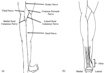
Figure 1
(a) Anatomic illustration of the sural nerve, (b) illustration of the
course of the sural nerve.
Figure 2
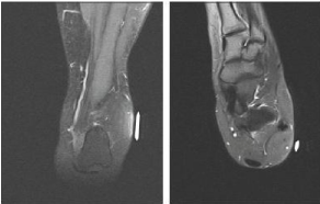
Figure 2
Coronal and saggital images of the ankle demonstrating the
subcutaneous lipoma.
Figure 3
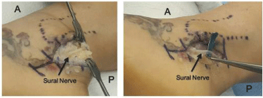
Figure 3
Surgical dissection revealing the sural nerve entrapped in the
lipoma.
Discussion
The sural nerve courses subcutaneously in the posterolateral aspect of the leg, passing over the
lateral edge of the Achilles tendon approximately 10 cm proximal to the calcaneus (Figure 1b).
Distally it lays 1-1.5 cm posterior to the posterior border of the lateral malleolus, giving rise to the
lateral calcaneal nerve and terminating at the lateral dorsal cutaneous nerve. 1 it further divides at the base of the fifth metatarsal forming the dorsal and plantar branches.
The dorsal branch supplies the dorsal digital nerve providing
sensation to the lateral side of fifth toe. It provides anastomoses to
other digital nerves to supply the medial border of the fifth and lateral
border of the fourth toe [2,4].
Cahill et al. [1] described the sural nerve as arising from the
combination of MSCN and LSCN in 80% of cases, and as a direct
continuation of MSCN in 20% of cases. The site of union of the
MSCN and LSCN varies as well, forming anywhere from 11-20 cm
proximal to the lateral malleolus [3]. Cahill et al. [1] described the
average length of the sural nerve to be 11-20 cm, with its diameter
ranging from 2.5 to 4.0 mm proximally and 2.0 to 3.0 mm distally;
and a central nutrient artery and vein.1 Coert et al. [5] described this
formation occurring in the popliteal fossa in approximately 12% of
cases, and in lower third of leg in 84% of cases.
Percutaneous sutures should be avoided in the lateral half of
Achilles tendon repairs due to its close proximity to the sural nerve
[3,6,7]. Hockenbury et al. [6] described a 60% incidence of sural
nerve injury during percutaneous repair of cadaveric Achilles tendon.
Non-surgical injury can occur as a result of entrapment by hematoma
as well as following Achilles tendon rupture [8,9]. Traction injury to
the sural nerve can occur as a result of Achilles rupture, which when
intact acts as a checkrein to prevent nerve traction in physiologic
conditions [8]. O’Brien et al. [10] described sural nerve entrapment
by scar tissue after rupture of the gastrocnemius muscle. EMG studies showed latency in the sural nerve distribution along with reduction
of sensory nerve action potential amplitude. MRI revealed abnormal
signal in the gastrocnemius consistent with a subacute hematoma.
Surgical exploration revealed encasement of the sural nerve in scar
tissue underneath deep fascia at the level of the gastrocnemius. The
release of the nerve resulted in restoration of sensation at the 3
month follow-up period [10]. Trevino et al. [11] reported on three
cases in which an avulsion fracture of the base of the fifth metatarsal
resulted in sural nerve entrapment. Sural nerve function was restored
following removal of the fragments causing entrapment [11].
Sural nerve entrapment can occur from direct traumatic
contusions to the nerve, fractures of the calcaneus, fifth metatarsal,
posterolateral process of the talus, or from os perineum with the most
common site of entrapment being the fibrous arcade of the Achilles
musculotendinous junction [2,4].
Entrapment can lead to chronic neuropathic pain and diagnosis
is based on clinical examination. A thorough history and physical
examination is important to rule out exertional compartment
syndrome and popliteal artery entrapment syndrome [12].
Neuropathy can present as constant, sharp, achy pain unresponsive
to analgesics, dysesthesias, associated with numbness and tingling in
the nerve distribution [13,14]. Characteristic signs in patients
with sural nerve entrapment includes tenderness in the middle part
of the calf, lateral to the musculotendinous junction of the Achilles
tendon; with a positive tinel's sign [12,14]. In cases of long-standing
nerve entrapment EMG studies show a decrease in nerve conduction
velocity and reduction in sensory potential amplitudes, however,
these studies can be negative [12]. Conservative treatment for
neuropathic pain includes using tricyclic antidepressants, serotonin
and norepinephrine reuptake inhibitors, and calcium channel
alpha2-delta agonists [13]. Todorov et al. [13] reported complete
relief of pain following pulsed radiofrequency for neuropathic Sural
nerve pain. Fabre et al. [12] demonstrated sural nerve entrapment
by an increase in sural muscle mass and development of local
fibrous scar tissue. Thirteen patients (18 limbs) were retrospectively
analyzed with calf tenderness along the course of the sural nerve
distribution. EMG studies were positive in 10 patients. After failure of
nonoperative treatment, patients underwent neurolysis with release
of the superficial sural aponeurosis and fibrous band in which the
nerve traversed. Excellent results were reported in 9 limbs, good in 8
limbs, and fair in 1 limb after 14 months follow-up [15].
Conclusion
The patient presented with a lipoma encasing the sural nerve and recovered well following its removal. There is a lack of literature on the incidence of sural nerve lipomas and its manifestations. The nerve arises as a compilation of the LSCN and MSCN. It courses posterolaterally about the ankle to provide sensation to the posterolateral aspect of the distal third of the leg and the lateral aspect of the fourth and fifth digits. Injuries to the sural nerve can be a complication of Achilles tendon repair or result of entrapment from a mass or fractures and lead to paresthesias or neuropathic pain.
References
- Ortigiiela ME, Wood MB, Cahill DR. Anatomy of the sural nerve complex. J Hand Surg . 1987; 12: 1119-1123.
- Chou LB. Orthopaedic Knowledge Update: Foot and Ankle 5. Am Acad Orthop Surg; 2014.
- Webb J, Moorjani N, Radford M. Anatomy of the sural nerve and its relation to the Achilles tendon. Foot Ankle Int. 2000; 21: 475-477.
- Gould JS. Preface. Nerve problems of the lower extremity. Foot Ankle clinic. 2011; 16: 11-12.
- Coert JH, Dellon AL. Clinical implications of the surgical anatomy of the sural nerve. Plast Reconstr Surg. 1994; 94: 850-855.
- Hockenbury RT, Johns JC. A biomechanical in vitro comparison of open versus percutaneous repair of tendon Achilles. Foot Ankle. 1990; 11: 67-72.
- Klein W, Lang DM, Saleh M. The use of the Ma-Griffith technique for percutaneous repair of fresh ruptured tendo Achillis. Chir Organi Mov. 1991; 76: 223-228.
- Fletcher MDA, Warren PJ. Sural nerve injury associated with neglected tendo Achilles ruptures. British J Sports Med. 2001; 35: 131-132.
- Fitzgibbons RE, Hefferon J, Hill J. Percutaneous Achilles tendon repair. Am J Sports Med. 1993; 21: 724-727.
- Bryan BM, Lutz GE, O'Brien SJ. Sural nerve entrapment after injury to the gastrocnemius: A case report. Arch Phys Med Rehabil. 1999; 80: 604-606.
- Gould N, Trevino S. Sural Nerve Entrapment by Avulsion Fracture of the Base of the Fifth Metatarsal Bone. Foot Ankle Int. 1981; 2: 153-155.
- Fabre T, Montero C, Gaujard E, Gervais-Dellion F, Durandeau A. Chronic Calf Pain in Athletes Due to Sural Nerve Entrapment: A Report of 18 Cases. Am J Sports Med. 2000; 28: 679-682.
- Todorov L. Pulsed radiofrequency of the sural nerve for the treatment of chronic ankle pain. Pain physician. 2011; 14: 301-304.
- Paraskevas GK, Natsis K, Tzika M, Ioannidis O. Fascial entrapment of the sural nerve and its clinical relevance. Anat Cell Biol. 2014; 47: 144-147.
- Fabre T, Montero C, Gaujard E, Gervais-Dellion F, Durandeau A. Chronic calf pain in athletes due to sural nerve entrapment. A report of 18 cases. Am J Sports Med. 2000; 28 : 679-682.