Case Report
Thyrotoxicosis due to Atypical Subacute Thyroiditis Scintigraphicaly Presented as Toxic Adenoma – Case Report
Željka Aleksić1*, Aleksandar Aleksić2 and Nenad Ristović3
1Department of Nuclear Medicine, Health Center Zaječar, Serbia
2Department of Internal Medicine, Health Center Zaječar, Serbia
3Department of Infectious Disease, Health Center Zaječar, Serbia
*Corresponding author: Željka Aleksić, Health Center Zaječar, Nuclear medicine Service, Nikole Pašića 88/3, 19 000 Zaječar, Serbia
Published: 17 Nov, 2016
Cite this article as: Aleksić Ž, Aleksić A, Ristović N.
Thyrotoxicosis due to Atypical Subacute
Thyroiditis Scintigraphicaly Presented
as Toxic Adenoma – Case Report. Ann
Clin Case Rep. 2016; 1: 1183.
Abstract
Subacute thyroiditis (SAT), also known as DeQuervain's or granulomatous thyroiditis is an inflammatory condition of the thyroid gland, which is usually fully affected. Severe pain and tenderness in the thyroid bed occur typically and are followed by weakness, fatigue, pain in muscles and joints, and light to moderate fever and symptoms of thyrotoxicosis - nervousness, sweating and rapid heart beat and trembling. Temporary elevated serum thyroxin, suppressed TSH and elevated serum markers of inflammation in the acute phase are pathognomonic for the SAT, with diffusely absent binding of radioactive iodine or technetium-pertechnetate in the thyroid gland due to thyrocytes destruction. Rare cases of atypical SAT - painless or with minimal pain, SAT limited to one thyroid lobe or focal thyroiditis, are reported in literature. We report a rare case of a minimally painful SAT associated with functional adenoma in the right lobe of the thyroid gland, which was, in thyrotoxic, acute phase, scintigraphicaly presented as toxic adenoma, in fact representing a functional adenoma tissue unaffected by destructive thyroiditis.
Keywords: Atypical subacute thyroiditis; Autonomously functioning thyroid nodule; Toxic adenoma
Introduction
Subacute thyroiditis (SAT), also known as DeQuervain's or granulomatous thyroiditis is an inflammatory condition of the thyroid gland, which is usually affected as a whole. It can last from several weeks to several months, probably of viral origin, also occasionally described as epidemic illness, resolve spontaneously, and it can recur. The most commonly affects women between the fifth and sixth decades of life. Typically it presents with severe pain and tenderness in the thyroid bad followed by weakness, fatigue, pain in muscles and joints, light to moderate fever and symptoms of thyrotoxicosis - anxiety, sweating, rapid heart beat and trembling. Rare cases of atypical SAT: painless or with minimal pain, SAT limited to one lobe of the thyroid, or focal thyroiditis are described in literature as well [1].
Case Presentation
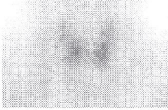
Seventy two-year-old female patient was hospitalized at the Infectious diseases department with fever of unknown origin and fatigue, which lasts about a month back. In the personal history there was a high blood pressure treated with ACE inhibitor and multiple sclerosis. Palpatory finding of the thyroid gland on admission was normal, and markers of inflammation, fibrinogen, and CRP were positive. The treatment with antibiotics for suspected urinary infection has been started, due to a finding of a positive urine culture. Given that the pain persisted, the tenth day of hospitalization thyroid hormones analyses were done, showing increased free fraction of thyroid hormones and suppressed TSH, with, still elevated fibrinogen. At this point, the patient was clinically moderately thyrotoxic with palpable, uneven, movable on swallowing thyroid gland, with slightly painful left lobe. On echosonography, thyroid gland was slightly diffusely enlarged, diffusely very heteroehogenous, predominantly hypoechogenic, with predominantly isoechogenic nodal formation distally in the right lobe, with diameter around 15,8x12,5x15,8 mm. Pertechnetate scan indicated the existence of a functional tissue in the lower half of the right lobe, with total suppression of surrounding thyroid tissue (Figure 1a). Thyroid scintigraphy with methoxy-isobutyl-isonitrile (MIBI) was performed and showed slightly decreased and inhomogeneous uptakewith the parts of more intensive uptake into the lower parts of both lobes (Figure 2). Glucocorticoid therapy was started with a gradually decreasing doses on five days, with a total length of about a month, along with monitoring of thyroid status. Clinical and biochemical thyroid status normalized about 6 weeks after diagnosis (Table 1). Control echosonography about 3 months after diagnosis, showed the existence of slightly hypoehoic nodule in the distal part of right lobe with diameter around 16x9x12 mm, with moderately heteroechogenous parenchyma outside the nodule. Control pertechnetate scan confirmed the existence of functioning adenoma in the distal part of right lobe and the recovery of uptake in the surrounding tissue of the thyroid gland (Figure 1b). The last checkup, about a year from the acute phase showed normal functional thyroid status (Table 1), echosonographically, finding was similar to the previous one, and on a thyroid pertechnetate scan functional adenoma with slightly decreased uptake in the surrounding tissue was present (functional adenoma of the right lobe) (Figure 1c).
Figure 1
Figure 1
Echosonographic findings and pertechnetate thyroid scan in a patient during follow-up
(a) the 1st visit. (b) Four months after 1st visit. (c) One year after 1st visit.
Table 2
Table 1
Findings of serum biochemical indicators of thyroid status and markers of inflammation as well as clinical status in patient during follow-up. EU-clinically
euthyroid; MB-palpable, minimally painful thyroid; BB – painless, non-palpable thyroid.
Figure 2
Discussion and Conclusion
The cause of subacute thyroiditis is rarely possible to determine.
Given that it often occurs after a respiratory infection, it is assumed
that it causes infectious agent. In some patients the serological analysis
or cultured thyroid tissue sample confirmed the presence of the virus
mumps, and recorded the occurrence of the epidemic occurrence
[2]. Other viruses that have been isolated in some patients, including
measles virus, influenza virus, H1N1 influenza virus, adenovirus,
Epstein-Barr virus, coxsackie virus B4, and even cytomegalo virus
[3-10]. It is believed that there is a genetic pre disposition to an
inflammatory response to infection with different thyroid viruses [10]. There are cases of subacute thyroiditis described in patients
treated with immunomodulatory therapy (interleukin 2, TNF-α
and interferon-γ) [11] as well as in patients after administration of
contrast media for radiological tests [12].
SAT is rare in children [13]. Several times more common in
women than in men, usually from the third to fifth decade of life
[14,15]. It is rare in pregnancy [16]. The incidence of the disease is
about five times lower than the incidence of Graves' disease [1].
It is typically presented with severe pain and tenderness in the
thyroid bad, which can spread to the jaw or ear, accompanied by
weakness, fatigue, pain in muscles and joints and light to moderate
fever, and symptoms of thyrotoxicosis - anxiety, sweating, rapid heart
and trembling. Difficulty with swallowing, even transient vocal cord
paresis were described also [17]. Thyroid gland is usually enlarged,
firm and painfully sensitive to palpation. The symptoms may
culminate in the third and fourth days of the onset of the disease, and
then gradually weaken and disappear in one week. However, the most
common symptoms develop gradually over one to two weeks, and
in the next 3-6 weeks after fluctuating weight and representation. In
some patients, during many months after of the onset of the disease,
many exacerbations of symptoms may occur until full recovery [1,18].
There are rare cases of atypical SAT described: painless, or with
minimal pain [19], SAT limited to one thyroid lobe, or focal thyroiditis
[20,21], and the thyroid storm caused by subacute thyroiditis as well
[22]. In the available literature we found one case report of SAT
coexisting with autonomously functioning nodule, similar to our
own described case [23].
Recovery after subacute thyroiditis is accompanied by transient hypothyroidism in about quarter of patients, but in less than 10% it may result in permanent hypothyroidism [24].
Pathognomonic for the SAT is temporary elevated serum thyroxin
level, suppressed TSH and elevated erythrocyte sedimentation rate
and other serum markers of inflammation (CRP, fibrinogen) in the
acute phase, with diffusely absent uptake of radioactive iodine or
technetium-pertechnetate in thyroid gland because of the destruction
thyrocytes. The serum concentration of thyroglobulin is increased
for the same reason, while the anti-thyroglobulin antibody and anti-
TPO antibody levels are usually negative [1]. In half of the patients
the increase of liver enzymes in the serum may be found, which may
be persist for up to several months [25].
Pertechnetate thyroid scan is cheap and convenient diagnostic
method to confirm SAT, where there is an absent uptake of radiotracer
due destructive process in the thyroid [26]. Thyroid scintigraphy with
fat soluble radiopharmaceuticals, such as MIBI or tetrofosmin may
in acute, thyrotoxic phase of the disease, show increased uptake of
the tracer in the thyroid, probably as a result of hyperemia caused
by inflammation [27,28]. In our experience in the present case, the
binding of MIBI in thyroid in the acute phaseis rather impaired than
enhanced, which can be interpreted as a result of destructive changes
in the thyroid also, knowing that MIBI is taken by viable cells. In the
acute, thyrotoxic phase, color flow Doppler sonography (CFDs) of
thyroid can be helpful in the differential diagnosis to other thyrotoxic
states (autoimmune or autonomous hyperthyroidism), because in
SAT there is absent CD signal in the affected thyroid [29]. Standard
gray scale sonography shows characteristic irregular hypoechogenic
fields of affected thyroid parenchyma, whose extensiveness is usually
a measure of severity of clinical manifestations [30]
.
Conditions which may be similarly clinically manifested as SAT
and come into consideration as a differential diagnosis entities are
pharyngitis [31], temporal arteritis [32], pain in the jaw teeth of origin
[33], bleeding in thyroid cysts [22], less often Hashimoto's thyroiditis.
In atypical, painless or minimally painful cases SAT, this should be
distinguished from the autoimmune painless thyroiditis, lymphocytic
thyroiditis variants, in which no systemic inflammatory signs and
symptoms are present [34]. Focal SAT can mimic suppurative
thyroiditis, and thyroid cancer as well [29].
The therapeutic approach depends on the severity of the clinical
picture. In some patients, there is no need for any medical therapy.
Some times it is necessary to apply non-steroidal anti-inflammatory
drugs and aspirin to reduce pain. In cases of severe symptoms glucocorticoid therapy could be indicated [35]. It is usually given in
a single daily dose of 40 mg of prednisone and at 7 days the dose
is gradually reduced by 5 mg for 6 weeks. An oral therapy with
cholecystography agents (sodium ipodate or sodium iopanoate) were
reported as safe and effective in controlling thyrotoxicosis induced
by subacute thyroiditis [36]. In the case of the development of
permanent hypothyroidism after an episode of SAT, levothyroxine
substitution is indicated.
We present an unusual case of a minimum painful SAT
associated with functional adenoma in the right lobe of the thyroid
gland, which scintigraphy presented as a toxic adenoma in thyrotoxic
phase, and in fact was a functional adenoma tissue unaffected with
destructive thyroiditis. On the standard sonography adenoma tissue
was more echogenic than the surrounding thyroid parenchyma,
which showed a typical heterogeneous hypoechoic appearance, and
the MIBI scan showed moderately intense tracer binding in the
thyroid gland. Serum markers of inflammation were typically high,
with suppressed TSH and elevated serum thyroxin in the acute phase,
with a gradual normalization during follow-up, but without transient
hypothyroid phase, probably as a consequence of the existence of
regional autonomy. Pertechnetate scan during follow-up showed the
functional recovery of the previously affected gland parenchyma with
destructive thyroiditis.
References
- Rupendra T. Shrestha, Hennessey J. Acute and Subacute, and Riedel’s Thyroiditis. 2015.
- Parmar RC, Bavdekar SB, Sahu DR, Warke S, Kamat, JR. Thyroiditis as a presenting feature of mumps. Pediatr Infect Dis J. 2001; 20: 637-638.
- Dimos G, Pappas G, Akritidis N. Subacute thyroiditis in the course of novel H1N1 influenza infection. Endocrine. 2010; 37: 440-441.
- Volta C, Carano N, Street ME, Bernasconi S. Atypical subacute thyroiditis caused by Epstein-Barr virus infection in a three-year-old girl. Thyroid. 2005; 15: 1189-1191.
- Satoh M. Virus-like particles in the follicular epithelium of the thyroid from a patient with subacute thyroiditis (deQuervain’s). Acta Pathol Jpn. 1975; 25: 499-501.
- Engkakul P, Mahachoklertwattana P, Poomthavorn P. de Quervain thyroiditis in a young boy following hand-foot-mouth disease. Eur J Pediatr. 2011; 170: 527-529.
- Luotola K, Hyoty H, Salmi J, Miettinen A, Helin H, Pasternack A. Evaluation of infectious etiology in subacute thyroiditis–lack of association with coxsackievirus infection. APMIS. 1998; 106: 500-504.
- Mori K, Yoshida K, Funato T, Ishii T, Nomura T, Fukuzawa H, et al. Failure in detection of Epstein-Barr virus and cytomegalovirus in specimen obtained by fine needle aspiration biopsy of thyroid in patients with subacute thyroiditis. Tohoku J Exp Med. 1998; 186: 13-17.
- Espino Montoro A, Medina Perez M, Gonzalez Martin MC, Asencio Marchante R, Lopez Chozas JM. Subacute thyroiditis associated with positive antibodies to the Epstein-Barr virus. An Med Interna. 2000; 17: 546-548.
- Al Maawali A, Al Yaarubi S, Al Futaisi A. An infant with cytomegalovirusinduced subacute thyroiditis. J Pediatr Endocrinol Metab. 2008; 21: 191- 193.
- Amenomori M, Mori T, Fukuda Y, Sugawa H, Nishida N, Furukawa M, et al. Incidence and characteristics of thyroid dysfunction following interferon therapy in patients with chronic hepatitis C. Intern Med. 1998; 37: 246-252.
- Calvi L, Daniels GH. Acute thyrotoxicosis secondary to destructive thyroiditis associated with cardiac catheterization contrast dye. Thyroid. 2011; 21: 443-449.
- Ogawa E, Katsushima Y, Fujiwara I Iinuma K. Subacute thyroiditis in children: patient report and review of the literature. J Pediatr Endocrinol Metab. 2003; 16: 897-900.
- Fatourechi V, Aniszewski JP, Fatourechi GZ, Atkinson EJ, Jacobsen SJ. Clinical features and outcome of subacute thyroiditis in an incidence cohort: Olmsted County, Minnesota, study. J Clin Endocrinol Metab. 2003; 88: 2100-2105.
- Qari FA, Maimani AA. Subacute thyroiditis in Western Saudi Arabia. Saudi Med J. 2005; 26: 630-633.
- Anastasilakis AD, Karanicola V, Kourtis A, Makras P, Kampas L, Gerou S, et al. A case report of subacute thyroiditis during pregnancy: difficulties in differential diagnosis and changes in cytokine levels. Gynecol Endocrinol. 2011; 27: 384-390.
- Dedivitis RA, Coelho LS. Vocal fold paralysis in subacute thyroiditis. Braz J Otorhinolaryngol. 2007; 73: 138.
- Benbassat CA, Olchovsky D, Tsvetov G, Shimon I. Subacute thyroiditis: clinical characteristics and treatment outcome in fifty-six consecutive patients diagnosed between 1999 and 2005. J Endocrinol Invest. 2007; 30: 631-635.
- Daniels GH. Atypical subacute thyroiditis: preliminary observations. Thyroid. 2001; 11: 691-695.
- Nakamura S, Saio Y, Ishimori M. Recurrent Hemithyroiditis: A case report. Endocr J. 1998; 45: 595-600.
- Sari O, Erbas B, ErbasT. Subacute thyroiditis in a single lobe. Clin Nucl Med. 2001; 26: 400-401.
- Swinburne JL, Kreisman SH. A rare case of sub acute thyroiditis causing thyroid storm Thyroid. 2007; 17: 73-76.
- Liel Y. The survivor: association of an autonomously functioning thyroid nodule and subacute thyroiditis. Thyroid. 2007; 17: 183-184.
- Nishihara E, Amino N, Ohye H, Ota H, Ito M, Kubota S, et al. Extent of hypoechogenic area in the thyroid is related with thyroid dysfunction after sub acute thyroiditis. J Endocrinol Invest. 2009; 32: 33-36.
- Matsumoto Y, Amino N, Kubota S, Ikeda N, Morita S, Nishihara E, et al. Serial changes in liver function tests in patients with subacute thyroiditis. Thyroid. 2008; 18: 815-816.
- Intenzo CM, Park CH, Kim SM, Capuzzi DM, Cohen SN, Green P. Clinical, laboratory, and scintigraphic manifestations of subacute and chronic thyroiditis. Clin Nucl Med. 1993; 18: 302-306.
- Hiromatsu Y, Ishibashi M, Miyake I Nonaka K. Technetium-99m tetrofosmin imaging in patients with subacute thyroiditis. Eur J Nucl Med. 1998; 25: 1448-1452.
- Hiromatsu Y, Ishibashi M, Nishida H, Kawamura S, Kaku H, Baba K, et al. Technetium-99 m sestamibi imaging in patients with subacute thyroiditis. Endocr J. 2003; 50: 239-244.
- Sun Young Park, Eun-Kyung Kim, Min Jung Kim, Byung Moon Kim, Ki Keun Oh, Soon Won Hong, et al. Ultrasonographic characteristics of subacute granulomatous thyroiditis. Korean J Radiol. 2006; 7: 229-234.
- Omori N, Omori K Takano K. Association of the ultrasonographic findings of subacute thyroiditis with thyroid pain and laboratory findings. Endocr J. 2008; 55: 583-588.
- Janssen OE. Atypical presentation of subacute thyroiditis. Dtsch Med Wochenschr. 2011; 136: 519-522.
- Cunha BA, Chak A, Strollo S. Fever of unknown origin (FUO): de Quervain’ssubacute thyroiditis with highly elevated ferritin levels mimicking temporal arteritis (TA). Heart Lung. 2010; 39: 73-77.
- Tesfaye H, Cimermanova R, Cholt M, Sykorova P, Pechova M, Prusa R. Subacute thyroiditis confused with dental problem. Cas Lek Cesk. 2009; 148: 438-441.
- Woolf PD. Transient painless thyroiditis with hyper-thyroidism: a variant of lymphocytic thyroiditis?. Endocr Rev. 1980; 1: 411-420.
- Volpe R. The management of subacute (DeQuervain’s) thyroiditis. Thyroid. 1993; 3: 253-255.
- Martinez DS, Chopra IJ. Use of oral cholecystography agents in the treatment of hyperthyroidism of subacute thyroiditis. Panminerva Med. 2003; 45: 53-57.


