Clinical Image
Large Myoma with Mixoid Degeneration, Fast Growing Fibroid or Uterine Sarcoma
Begoña Díaz de la Noval*
Department of Gynecological Oncology, La Paz University Hospital, Spain
*Corresponding author: Begoña Díaz de la Noval, Department of Gynecological Oncology, European Society of Gynaecology Oncology (ESGO) Fellow, Gynecology Oncology Unit. La Paz University Hospital, Paseo de la Castellana 261, 28046-Madrid, Spain
Published: 10 Oct, 2016
Cite this article as: Díaz de la Noval B. Large Myoma with
Mixoid Degeneration, Fast Growing
Fibroid or Uterine Sarcoma. Ann Clin
Case Rep. 2016; 1: 1150.
Abstract
Introduction and Aim: There is an increased risk of uterine sarcoma in women with an enlarged and active growing myoma. Our aim is to make a short report of a case based on clinical images.
Clinical Image: A 50-year-old woman with a giant mass that occupied the whole abdominal cavity,
with exophytic growth through the vagina. Imaging studies described a solid tumor of 27 x 20 x 30
cm suggestive of myoma or sarcoma; right hydronephrosis and inferior vena cava compression.
No peritoneal involvement. Total abdominal hysterectomy and bilateral salpingo-oophorectomy
by open surgery and an exhaustive abdominal cavity review were performed. Histological analysis
confirmed a 32 x 27 cm myoma with myxoid degeneration, no malignant cells. The patient had a
great recovery with no urinary or vascular complications.
Discussion: There is high-risk of degeneration to a sarcoma in a large uterus, fast growing
myomas and perimenopause, due to the phase of change or more marked hormonal alteration.The
gynecologist should monitor these patients with more frequency, especially if the myoma grows
rapidly or has changed.
Conclusion: Active or big myomas are associated with occult uterine sarcoma; however, this
criterion is not enough to allow a preoperative identification in all patients in the same condition.
Meanwhile, an exhaustive abdominal cavity review is recommended to be performed during surgery.
Future studies to further development of a Score in cases of suspected malignancyare needed.
Clinical Image
A 50-year old woman with a solid tumor of 27 x 20 x 30 cm that occupies the abdomen,
with exophytic growth through the vagina; suggestive or sarcoma [1]. The patient complained of
progressive abdominal distension for more than a year ago. She blamed against weight increase
with the onset of menopause. The patient complained about a right hydronephrosis and vena cava
compression (Figure 1), due to voiding difficult and moderate edema in legs. The radiological test shows a big abdominal mass depending on the uterus and no signs of carcinomatosis.
We suspected a high risk of uterine sarcoma or other malignancy [2], so we decided to perform
a total abdominal hysterectomy and bilateral salpingo-oophorectomy by laparotomy (Figure 2).
Final histology analysis confirmed a giant uterine myoma with myxoid degenerated [3,4], no signs of malignancy [5]. The patient had a great recovery with no urinary or vascular complications. No postoperative complications. Discharged without incident. Follow-up free of disease.
Figure 1
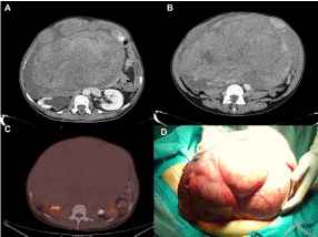
Figure 1
(a-c) CT and PET-CT without peritoneal disease or distant metastases. (d) Enlarged uterus of
33x32x19cm due to a cervical large myoma of 32x27cm.
Figure 2
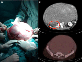
Figure 2
(a) Giant myoma. (b,c) Right hydronephrosis and inferior vena cava compression.
Ethical Statement
Written informed consent of the patient for publication has been obtained.
References
- Cho HY, Kim K, Kim YB, No JH. Differential diagnosisbetween uterine sarcoma and leiomyoma using preoperative clinical characteristics. J Obstet Gynaecol Res. 2016; 42: 313-318.
- Yamashiro T, Gibo M, Utsunomiya T, Murayama S. Huge uterine leiomyoma with adenomyotic cysts mimicking uterine sarcoma on MR imaging. Radiat Med. 2007; 25: 127- 129.
- Takano Y, Morimura Y, Yamada H, Yanagida K, Sato A, Suzuki O, et al. Myxoidleiomyosarcoma of the uterus. Fukushima J Med Sci. 2000; 46: 41- 47.
- Kim HS, Yoon G, Jung YY, Lee YY, Song SY. Fibromyxoid variant of endometrial stromal sarcoma with atypical bizarre nuclei. Int J Clin Exp Pathol. 2015; 8: 3316-3321.
- Novetsky AP, Powell MA. Management of sarcomas of the uterus. Curr Opin Oncol. 2013; 25: 546-552.