Case Report
Painful Early Cancerous Lesion Needs Evaluation: A Case Report
Hitesh Verma*, Ananya Soni, Kapil Sikka, David Victor Kumar Irugu, Alok Thakar
1Department of Otorhinolaryngology and Head & Neck Surgery, All India Institute of Medical Sciences, India
*Corresponding author: Hitesh Verma, Department of Otorhinolaryngology and Head & Neck Surgery 4057, ENT Office, 4th Floor, Teaching Block, All India Institute of Medical Sciences, New Delhi, India
Published: 03 Oct, 2016
Cite this article as: Verma H, Soni A, Sikka K, Irugu DVK,
Thakar A. Painful Early Cancerous
Lesion Needs Evaluation: A Case
Report. Ann Clin Case Rep. 2016; 1:
1149.
Abstract
Several drugs have previously been implicated to cause acute pancreatitis. We report the rare case of codeine induced acute pancreatitis. A 43-year-old woman developed the symptoms and clinical signs of acute pancreatitis 90 minutes after the ingestion of codeine phosphate. The diagnosis was proved both biochemically and radiologically which excluded other potential causes. The likely underlying pathophysiology is codeine-induced spasm of the sphincter of oddi.
Keywords: Lip cancer; Perineural invasion; MRI
Introduction
Lips are most common site for head and neck malignant lesions. The lip lesion by its prominent location is generally detected in early stage. The stage and location of tumor are influential factor for cancerous pain. The tumor generates pain by triggering infection and inflammation by crushing and infiltration. Early lesion can involve mental nerve and mandible by direct extension, perineural invasion and lymphatic spread into mental foramen. The perineural extension of disease is generally asymptomatic in early stage but alarm rings when pain, paresthesia, numbness are the presetting symptom. Perineural disease remains a diagnostic, prognostic and therapeutic challenge for the multidisciplinary team approach [1]. The perineural invasion associated with high chances of loco regional recurrence and affect survival if missed [2]. We are highlighting here a rare case of lower lip malignancy in its central part with pain in the region of mental nerve distribution and its management.
Case Presentation
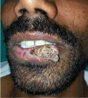
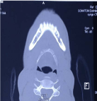
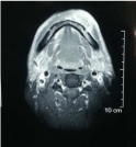
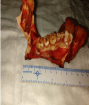
Thirty six year male patient presented with 6 years history of non healing ulcer on lower lip. The clinical examination revealed 3×2 cm ulcer on left side of lower lip extending from vermilion border to 3 cm from gingivolabial sulcus in its antero-posterior direction (Figure 1). In horizontal direction, extension was from midline to 2 cm short of left commissural area. The local examination also showed 2×2 cm enlarged lymph node at left level 1b. The patient was posted for wide local excision and left side modified neck dissection. After admission patient was giving history of pain in the region of skin over mantum. This raised the suspiciousness of nerve involvement. The CECT showed widening of mental foramen and canal on left side mandible (Figure 2). The contrast MRI revealed thickness of mental nerve and mandibular nerve on left side till foramen ovale (Figure 3). The stage of tumor was changed from three to four and surgical plan was converted into wide local excision with segmental mandibulectomy and left MND with free fibular grafting (Figure 4). Intra-operative proximal part of mandibular nerve was send separately. The final histopathology report highlights the presence of perineural invasion of mantal nerve and mandibular nerve with disease free proximal end of mandibular nerve which was send separately in operation. The patient received post operative radiotherapy and he is under disease free regular follow up till date.
Figure 1
Figure 1
Clinical photograph revealing ulcer on left side lower lip ( 2 cm from commissure left side).
Figure 2
Figure 3
Figure 4
Discussion
The lip lesions diagnosed early and prognostically better among oral malignancies due to its prominent location. The usual presentation is non healing painless ulcer. Lower lip is more commonly involve then upper lip. The risk factors for mutation of oncogene are similar with other subside of oral cavity such as smoking, alcohol, UV radiation etc [3]. The bony invasion is most documented local cause of pain which was absent in our case. The causes of local pain are direct infiltration, presence of infection and by chemical reaction. The perineural invasion is a well-recognized clinicopathologic entity found in head and neck cancers. Perineural disease remains a diagnostic, prognostic and therapeutic challenge for the multidisciplinary team approach [4]. The nerves are important routes of tumor spread in H&N malignancies, yet the biology and prognostic implications of peri-neural tumor growth are not fully understood. On balance, the available evidence suggests that it is associated with an increased risk of loco-regional recurrence. MRI is the best imaging modality to detect tumor extent as we seen in our case. The suppression of high intensity fat signal at foramina on T1 weighted images is suggestive of tumor infiltration/ nerve involvement. The administration of contrast with fat suppression is facilitating better nerve evaluation [5]. The features of nerve infiltration on CT scan are widening of foramina, bony canal and increase thickness similar to our case [6].
Conclusion
The changes in symptomatology in early stage tumor need evaluation for better outcome and to explain prognosis.
References
- Nersesyan H, Slavin KV. Current aproach to cancer pain management: Availability and implications of different treatment options. Ther Clin Risk Manag. 2007; 3: 381-400.
- Garzino-Demo P, Zavattero E, Franco P, Fasolis M, Tanteri G, Mettus A, et al. Parameters and outcomes in 525 patients operated on for oral squamous cell carcinoma. J Craniomaxillofac Surg. 2016; 44: 1414-1421.
- Kumar M, Nanavati R, Modi TG, Dobariya C. Oral cancer: Etiology and risk factors: A review. J Cancer Res Ther. 2016; 12: 458-463.
- Jardim JF, Francisco AL, Gondak R, Damascena A, Kowalski LP. Prognostic impact of perineural invasion and lymphovascular invasion in advanced stage oral squamous cell carcinoma. Int J Oral Maxillofac Surg. 2015; 44: 23-28.
- Ong CK, Chong VF-H. Imaging of perineural spread in head and neck tumours. Cancer Imaging. 2010; 10: S92-S98.
- Ginsberg LE. Imaging of perineural tumor spread in head and neck cancer. Semin Ultrasound CT MR. 1999; 20: 175-186.


