Case Report
A Hairy Congenital Back Mass: Case Report and Literature Review
Klinger Gil1,5*, Levy David1, Konen Osnat2,5, Yakimov Maxim4 and Ben Amitai Dan3,5
1Department of Neonatology, Schneider Children’s Medical Center, Israel
2Department of Radiology, Schneider Children’s Medical Center, Israel
3Department of Dermatology, Schneider Children’s Medical Center, Israel
4Department of Pathology, Rabin Medical Center, Israel, Tel Aviv, Israel
5The Sackler School of Medicine, Tel Aviv University, Israel
*Corresponding author: Gil Klinger, Neonatal Intensive Care Unit, Schneider Children’s Medical Center of Israel, 14 Kaplan Street, Petah Tiqva 49202, Israel
Published: 03 Oct, 2016
Cite this article as: Klinger G, Levy D, Konen O, Yakimov
M, Ben Amitai D. A Hairy Congenital
Back Mass: Case Report and Literature
Review. Ann Clin Case Rep. 2016; 1:
1148.
Abstract
We present a female neonate born with an unusually located hairy back mass. There were no further
anomalies on physical examination. The mass was not discovered on routine prenatal sonography.
Complete surgical removal was achieved without complications and histological examination was
consistent with mature teratoma.
The differential diagnosis of back masses in the newborn and the classification and work-up of
neonatal teratomas are reviewed.
This case suggests that even though most teratomas are located in the sacroccocygeal region, prenatal
screening should not overlook the possibility of additional uncommon locations.
Keywords: Neonatal teratoma; Posterior thoracic mass; Congenital anomalies
Abbreviations
SCT: Sacrococcygeal Teratoma
Introduction
Congenital back masses are relatively common in the newborn period and are most commonly
located in the sacrococcygeal area in a midline position [1]. These masses may represent a congenital
anomaly of the spinal tract or a congenital tumor. When a back mass occurs in a location other than
the sacrococcygeal area, especially if it is not in a midline position, it may be overlooked because less
attention may be given to areas where masses are less likely to occur.
We present a case with an unusually located back mass and the diagnostic evaluation and
subsequent management.
Case Presentation
A female neonate was born at 37 weeks gestation, the second child of a healthy 44 year old
woman. Family history was unremarkable and there was no history of exposure to medications or
of substance abuse. Pregnancy follow-up was uneventful, with the exception of a growing uterine
myoma that required Cesarean delivery. Apgar scores at birth were 9 and 10 at 1 and 5 minutes,
respectively. After delivery a posterior mass was noted and the child was transferred to the Neonatal
Intensive Care Unit for further evaluation.
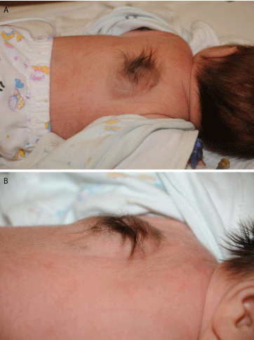
Initial physical examination showed a posterior thoracic mass, covered with hair (Figure
1).Vital signs were normal, no additional congenital abnormalities were noted and the neurological
exam was normal. Close examination of the mass showed that it was mostly located on the left
side of the thorax, but extended medially close to the midline. The mass was of a mixed solid and
cystic consistency and did not show any signs of inflammation. Chest X-ray showed a solid mass
with ossification at the level of T3-T6. Ultrasound showed a heterogenic mass with solid, cystic and
calcified areas and suggested possible invasion of the spinal cavity. Additional imaging including
head and abdominal ultrasound scans and a thoracic spine X-ray were normal. Blood chemistry and
complete blood count were normal as were β-human chorionic gonadotropin and alpha fetoprotein.
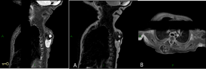
A magnetic resonance imaging scan (Figure 2) showed a 2.1 x 3.5 x 1.7 cm mass, located in the soft tissues of the back with solid and cystic consistency, extensive
calcifications and some fat tissue. It did not involve the muscles of the
upper back nor did it invade the spinal cavity. The spinal cord had a
normal signal and structure. The magnetic resonance scan concluded
that the mass was most likely a teratoma.
Surgery excision was performed on day 12 of life with no
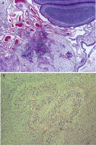
complications. Histological exam was consistent with a mature
teratoma (Figure 3).
The patient was discharged from hospital 4 days after the
procedure. Follow-up at 2 months showed normal development and
a normal neurological examination.
Figure 1
Figure 2
Figure 2
Magnetic resonance imaging of the thorax.
(a) Sagittal T1 WI F. (b) T2 WI of the back showing a complex mass with cystic component and a solid component with fat and calcifications and heterogeneous
enhancement following Gadolinium injection.
Discussion
Back masses in the newborn period are commonly located in the
sacrococcygeal area. These masses often represent neural tube defects,
or a sacrococcygeal teratoma (SCT). However, thoracic back masses
occurring in a non-midline position are most likely not related to the
spinal tract and have a different differential diagnosis.
The differential diagnosis of chest wall masses includes a wide
range of pathologies [1,2]. Midline masses may represent congenital anomalies of the spinal cord. Benign lesions include lipoma,
neurofibroma, lymphangioma, tufted angioma, hemangioma,
ganglioneuromaand mesenchymal hamartoma. Malignant lesions
include neuroblastoma, rhabdomyosarcoma and primitive
neuroectodermal tumors. Masses may also represent systemic diseases
such as leukemia, lymphoma and Langerhans cell histiocytosis. The
main clinical differential diagnoses considered in the case presented
were teratoma, congenital melanocytic nevus, meningocele,
spinal fusion abnormality with hemangioma or lipoma, plexiform
neurofibroma and ganglioneuroma. When evaluating the origin of a
congenital back mass its position, consistency, size, color, additional
skin findings, evolution with time and imaging findings should all
be considered. Imaging is diagnostic for spinal abnormalities and
often for teratomas, however for most of the above mentioned lesions
there are no diagnostic imaging characteristics. The mass identified in
our infant initiated close to the midline and extended laterally, thus
a neural tube defect and a teratoma were considered less likely. The
consistency of the mass was solid and cystic suggesting the possibility
of a teratoma. The color of the mass was somewhat bluish suggesting
a vascular component such as a hemangioma. The hair on the mass
was compatible with a teratoma or a neurofibroma [3].
Teratomas are a common form of Germ Cell Tumor and as all
germ layers are represented, its' cells may differentiate into any body
tissue including hair, teeth, fat, skin, muscle and others. Teratomas
are the most common tumor identified in the perinatal period, with
an incidence of about 1/20,000 live births [3]. The estimated incidence
of SCT, the most common teratoma, is 1 per 35,000 to 40,000 live
births [4,5]. SCT's have a female-to-male ratio of 4:1 [4,5]. Locations
where teratomas may occur are described in the Kiel Pediatric Tumor
Registry that includes 541 teratoma specimens. The most frequent
tumor sites are: the sacrococcygeal region (33.8%), the ovaries (31.2%)
and the testes (10.5%). Rare localizations include the mediastinum,
the retro peritoneum, the head and neck region as well as the central
nervous system [6].
Morbidity and mortality are usually low when the teratoma is
mature, diagnosed prenatally and with early surgical treatment [7].
Delay in diagnosis and treatment can lead to complications and
increased perinatal mortality. Patients with teratomas can have
coincident malformations in up to 18% of cases [8]. Follow up is
cardinal because the recurrence rate after complete resection of a
mature teratoma can be as high as 10-20% of patients [9]. The risk of
malignancy is about 10% in neonates and increases with age [9].
Teratomas are usually diagnosed on the basis cystic and solid
components identified by ultrasonography or MRI, either prenatally [10], often as early as the 1st trimester [10] or postnatally [11]. The
present case was preoperatively diagnosed as teratoma according
to clinical presentation and imaging studies. The diagnosis was
confirmed histologically, and was classified as mature solid and
cystic teratoma [12]. The main novelty in the case depicted above is
the unusual location of the teratoma. In a summary of 717 cases of
teratomas of infancy and childhood by Grosfeld and Billmire [13],
there was only one mature teratoma. However, this teratoma was
not identified in the neonatal period, was located in the scapular area
and not on the back, and was not close to the midline [14]. When a
mass compatible with teratoma is identified in utero or after birth in
areas not typical of teratoma, this diagnosis should still be taken into
account. The hairy nature of the mass should suggest the diagnosis of
a teratoma. The potential complications associated with birth of an
infant with a large posterior mass were avoided in the present report
as a cesarean delivery was performed.
We conclude that while performing prenatal screening, the
examiner must not overlook the possibility of a teratoma that is not
located in the sacral area. Hairy masses suggest a possible teratoma.
Figure 3
Figure 3
(a) Histology of back mass: the figure shows cerebellar tissue
and skin appendages with mature eccrine glands. (b) Glial tissue with basal
nucleus-like structure.
References
- Tibbs PA, James HE, Rorke LB, Schut L, Bruce DA. Midline hamartomas masquerading as meningomyeloceles or teratomas in the newborn infant. J Pediatr. 1976; 89: 928-933.
- La Quaglia MP. Chest wall tumors in childhood and adolescence. Sem Ped Surg. 2008; 17: 173-180.
- Na Ch, Song IG, Chung BS, Shin BS. Case of pigmented neurofibroma with hypertrichosis with no association to neurofibromatosis. J Dermatol. 2009; 36: 541-544.
- Varras M, Diakakis G, Monselas I, Akrivis CH. Prenatal diagnosis of a huge foetal immature sacrococcygeal teratoma: our experience of a rare case and review of the literature. OA Case Reports. 2013; 2: 10.
- Swamy R, Embleton N, Hale J. Sacrococcygeal teratoma over two decades: Birth prevalence, prenatal diagnosis and clinical outcomes. Prenat Diagn. 2008; 28: 1048-1051.
- Harms D, Zahn S, Gobel U, Schneider DT. Pathology and molecular biology of teratomas in childhood and adolescence. Klin Padiatr. 2006; 218: 296-302.
- Saygili-Yilmaz ES, Incki KK, Turgut M, Kelekci S. Prenatal diagnosis of type I sacrococcygealteratoma and its management. Clin Exp Obstet Gynecol. 2008; 35: 153-155.
- Altman RP, Randolph JG, Lilly JR. Sacrococcygeal teratoma: American Academy of Pediatrics surgical section survey-1973. J Pediatr Surg. 1974; 9: 389-398.
- Rescorla FJ, Sawin RS, Coran AG, Dillon PW, Azizkhan RG. Long-term outcome for infants and children with sacrococcygealteratoma: A report from the children's cancer group. J Pediatr Surg. 1998; 33: 171-176.
- Cay A, Bektas D, Imamoglu M, Bahadir O, Cobanoglu U, Sarihan H. Oral teratoma: a case report and literature review. Pediatr Surg Int. 2004; 20: 304-308.
- Adzick NS, Crombleholme TM, Morgan MA, Theresa M Quinn. A rapidly growing fetal Teratoma. Lancet. 1997; 349: 538.
- Grosfeld JL, Billmire DF. Teratomas in infancy and childhood. Curr Probl Cancer. 1985; 9: 1-53.
- Tapper D, Lack EE. Teratomas in Infancy and Childhood. A 54-Year Experience at the Children's Hospital Medical Center. Ann Surg. 1983; 198: 398-410.


