Case Report
A Rare Cause of Gastrointestinal Bleeding: Jejuneal Gastrointestinal Stromal Tumour
Ramazan Kuşaslan1, Talar Vartanoğlu1, Gülçin Hepgül1, Osman Bilgin Gulcicek1*, Yüksel Altınel1, Mehmet Öncü2, Yazgı Köy3, Şenay Yalçın3 and Fatih Çelebi1
1Department of General Surgery, Bagcilar Training and Research Hospital, Turkey
2Department of Radiology, Bagcilar Training and Research Hospital, Turkey
3Department of Pathology, Bagcilar Training and Research Hospital, Turkey
*Corresponding author: Osman Bilgin Gulcicek, Department of General Surgery, Bagcilar Training and Research Hospital, Bagcilar, Istanbul, 34200, Turkey
Published: 19 Sep, 2016
Cite this article as: Kuşaslan R, Vartanoğlu T,Hepgül G,
Gulcicek OB, Altınel Y, Öncü M, et
al. A Rare Cause of Gastrointestinal
Bleeding: Jejuneal Gastrointestinal
Stromal Tumour. Ann Clin Case Rep.
2016; 1: 1145.
Abstract
Gastrointestinal stromal tumors (GISTs) are the most common mesenchymal tumors of the gastrointestinal tract but are rarely malignant tumors of the digestive tract, accounting for 0.1% to 3.0% of all gastrointestinal neoplasms. They commonly arise from the stomach (40%-70%) and small intestine (20%-40%); other rare intestinal sites are the colon and rectum (5%-15%), and oesophagus (<5%). They can also rarely involve extraintestinal sites including the omentum, retroperitoneum, and mesentery (extra-intestinal GISTs). As a result of immunohistochemical and electronemicroscopical studies, GISTs are thought to be arised from the pacemaker cells of intestine known as Cajal cells. They are sometimes accompanied by symptoms, however in most cases are detected by chance. Complete resection is the primary treatment in the management of localized GISTs. Half of the patients have disease relapse in the first five years of surgery, and 5-year actuarial survival rate after surgery was reported as 54%. In the present case, a 70-year-old female patient presented to our emergency department with dizziness and blood in the stool. The patient was discharged in good overall condition.
Keywords: Gastrointestinal stromal tumor; Hemorrhage from gastrointestinal tract
Introduction
Gastrointestinal stromal tumors (GISTs) are the most common mesenchymal tumors of the
gastrointestinal tract but are rarely malignant tumors of the digestive tract, accounting for 0.1% to
3.0% of all gastrointestinal neoplasms [1]. The malignant potential of GIST is variable ranging from
small lesions with a benign behavior to fatal sarcomas. They commonly arise from the stomach
(40%-70%) and small intestine (20%-40%); other rare intestinal sites are the colon and rectum
(5%-15%), and oesophagus (<5%). They can also rarely involve extra intestinal sites including the
omentum, retroperitoneum, and mesentery (extra-intestinal GISTs) [2]. As a result of immune
histochemical and electromicroscopical studies, GISTs are thought to be arised from the pacemaker
cells of intestine known as Cajal cells. They are sometimes accompanied by symptoms, however
in most cases are detected by chance. Gastrointestinal stromal tumors (GISTs) are rare tumors
that may arise from any site of the GI tract and are generally associated with abdominal pain, GI
bleeding, or a palpable mass. However, a small intestinal GIST rarely causes hemorrhagic shock.
We here in report a case of hemorrhagic shock with excessive bleeding caused by anjejuneal GIST
that was managed by emergency surgery. The patient provided written informed consent for the
publication of this case report.Complete resection is the primary treatment in the management of
localized GISTs. Half of the patients have disease relapse in the first five years of surgery and 5-year
actuarial survival rate after surgery was reported as 54% [3].
In the present case, a 70-year-old female patient presented to our emergency department with
dizziness and blood in the stool. The patient was discharged in good overall condition.
Case Presentation
A 70-year-old female patient presented to the Department of Emergency of the Bagcilar
Research and Training Hospital with dizziness and blood in the stool. The patient's medical
history included treatment for hypertension by a local physician. The findings of the subsequent
physical examination were unremarkable, except for low blood pressure (100/60 mmHg) and mild
pallor of the conjunctiva. Laboratory data revealed severe anemia (hemoglobin, 7, 3 g/dl), INR: 1.33, WBC: 8200mm3, PLT: 276000mm3. Upper endoscopy revealed
no hemorrhagic lesion of the duodenum, stomach, or esophagus.
Colonoscopy revealed fresh blood with clotting discharged from the
proximal side of the ileocecal valve; hemorrhagic areaswere identified
at the colon or rectum. In the scintigraphical study, there was no
hemorrhagic focus.
The abdominal computed tomography (CT) revealed the 59 x
37 mm exophytic mass in the small intestine, The intestinal bleeding
continued, and the patient eventually developed hemorrhagic shock.
Hence, 2 units of red blood cells and 1 units of fresh-frozen plasma
were administered.Emergency partial resection of the jejunum tumor
which was 100 cm distaltothe ligament of Treitzwas performed. There
were no signs of lymphadenopathy, peritoneal dissemination, or liver
metastasis. The excised tumor (59mm x 37mm) exhibited ulcerative
mucosal changes. Sectioning of the tumor revealed a solid and
grayish white tissue. Histological examination of the excised tumor
revealed proliferation of spindle-shaped cells in the submucosa to the
subserosa of the ileum and a ruptured intratumoral artery at the tumor
surface (Figure 1). Immunohistochemical staining of the tumor was
positive for CD34, KIT and α-smooth muscle actin, but negative for
S-100 protein. The MIB-1 labeling index using Ki-67 was 1.0–5.0%.
The tumor size and the immunohistological findings supported the
diagnosis of a low-risk GIST of the jejunum. The patient had an
uneventful recovery, was discharged on postoperative day 7.
This case demonstrated the efficacy of the diagnosis of small
intestinal bleeding, and immediate emergency surgery should be
considered for cases of small intestinal GISTs with excessive bleeding.
Discussion
The clinical presentation of GISTs is variable and the most
frequent symptoms are abdominal pain, GI bleeding, or a palpable
mass. GISTs arise in any site, including the stomach, duodenum, small intestine, colon and rectum. However, with respect to small
intestinal GISTs, abdominal pain (35.5%) is the most frequent
symptom, while hemorrhagic shock (6.4%) is relatively rare. Owing
to limited information about the natural course and malignant
potential of GISTs less than 2 cm in size and the difficulty in targeting
and completely resecting such tumors, regular follow-ups rather
than resection have been recommended [4]. Clinical symptoms and
ailments related to the presence of a tumor largely depend on its
location and growth direction. Tumors with an outward pattern of
growth develop unnoticed, which is why they are not discovered in
the early stages. In this case, the patient developed hemorrhagic shock;
thus, upper endoscopy, colonoscopy, scintigraphy and abdominal
CT were performed to investigate the source of the hemorrhage. The
abdominal computed tomography (CT) revealed the 59mm x 37mm
exophytic mass in the small intestine. The most common clinical
indications of GISTs include obscure bleeding, abdominal pain,
and anemia. The first-line treatment of small intestinal GISTs with
excessive bleeding remains debatable. In the case, the CT located the
level of the bleeding and guided resection. In the present case, we
performed emergency partial resection of the jejunum, including the
GIST.
In conclusion, identify the source of bleeding in the small intestine
is more essential before the symptoms begin. Moreover, immediate
emergency surgery should be considered for cases of small intestinal
GISTs with excessive bleeding.
Acknowledgement
Present case was presented in 10th National Trauma and Emergency Congress, Antalya 2015, (poster presentation).
Figure 1
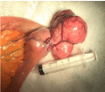
Figure 1
Median longitudinal groove with fissures arising on either side giving a fir tree appearance with
enlarged lunula and exfoliation of skin involving both thumb nails.
References
- Miettinen M, Sarlomo-Rikala M, Lasota J. Gastrointestinal stromal tumors: recent advances in understanding of their biology. Hum Pathol. 1999; 30: 1213-1220.
- Miettinen M, Majidi M, Lasota J. Pathology and diagnostic criteria of gastrointestinal stromal tumors (GISTs): a review. Eur J Cancer. 2002; 38: S39-S51.
- DeMatteo RP, Ballman KV, Antonescu CR, Corless C, Kolesnikova V, von Mehren M, et al. Long-term results of adjuvant imatinib mesylate in localized, high-risk, primary gastrointestinal stromal tumor: ACOSOG Z9000 (Alliance) intergroup phase 2 trial. Ann Surg. 2013; 258: 422-429.
- Fernández-Esparrach G, Sendino O, Solé M, Pellisé M, Colomo L, Pardo A, et al. Endoscopic ultrasound-guided fine-needle aspiration and trucut biopsy in the diagnosis of gastric stromal tumors: a randomized crossover study. Endoscopy. 2010; 42: 292-299.