Case Report
Adult Mediastinal Mesenteric Cyst: A Rare Mediastinal Tumor
Rezaeetalab F1*, Haghi Z2 and Dehestani V3
1Department of Pulmonology, Lung Disease Research Center, Mashhad University of Medical Sciences, Iran
2Department of Thoracic surgery, Mashhad University of Medical Sciences, Iran
3Lung Disease Research Center, Mashhad University of Medical Sciences, Iran
*Corresponding author: Fariba Rezaeetalab, Department of pulmonology , Lung Disease Research Center, Mashhad University of Medical Sciences, Mashhad, Iran
Published: 19 Sep, 2016
Cite this article as: Rezaeetalab F, Haghi Z, Dehestani V.
Adult Mediastinal Mesenteric Cyst: A
Rare Mediastinal Tumor. Ann Clin Case
Rep. 2016; 1: 1141.
Abstract
We report on a case of a mediastinal mesenteric cyst occurred in a 30-years-old woman was admitted
to the hospital due to chest pain of 3 months duration. Chest radiography revealed mediastinal
mass. Computed tomography (CT) of the chest showed heterogeneous mediastinal mass with
pleural effusion. The patient underwent open thoracotomy. Therefore, enterogenous cysts at ectopic
locations should be kept in mind.
Keywords: Mesenteric cyst; Enterogenous; Mediastinal; Mass
Introduction
Mesenteric cyst is one of the rare abdominal tumors. They usually arise from developmental abnormalities of the mesenteric lymphatics or from their traumatic rupture [1]. The lack of characteristic clinical features and radiological signs may result in great diagnostic difficulties. So they are discovered either accidentally during an abdominal radiological examination for another reason or during a laparotomy for the management of their complications such as torsion, rupture, haemorrhage of the cyst or adjacent organ obstruction [2-4]. Nearly 75% of mediastinal enterogenous cysts are recognized in the childhood. For unclear reasons, there is a two-to-one predilection for these cysts to be on the right side. Common symptoms are cough, dyspnea and occasionally stridor. The characteristic CT and MRI findings are identical to those of bronchogenic cysts apart from its wall which may be thicker and in near contact with the esophagus [3-8].
Case Presentation
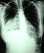
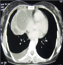
30-years-old woman was admitted to the hospital due to a chest pain of 3 months duration. Pain was often felt as a dull ache in the right hemi thorax. She did not have fever, cough, dyspnea, and hemoptysis or weight loss. She also did not report any arthralgia or any mucocutaneous, gastrointestinal or urogenital symptoms. She was a married housekeeper and lived with her husband and two children. She was not alcoholic or a smoker. Her general appearance was good and she did not seem ill. Her vital signs were as follow: BP = 120/80 mmHg, pulse rate = 86 per minute, respiratory rate=12 per minute, and temperature=37°C. Her physical examination was all normal including her lungs which were clear. Electrocardiography was also normal too. Blood indices, electrolytes urinalysis and renal function tests were normal. Her Erythrocyte Sedimentation (ESR) was 12 with Tuberculin skin test, acid fast stain, culture of gastric contents and bronchoalveolar lavage all negative for mycobacterium tuberculosis. Moreover, the hydatid agglutination test was 6 U/L (with upper limit of 15U/L). Chest- X-ray showed an opacity in the middle and lower zone of the right lung (Figure 1). Computed tomography (CT) of the chest showed a mass in the mediastinum (Figure 2). A Bronchoscopy was then performed with no abnormal findings. This was followed by the right thoracotomy. At the operation, a thick walled cyst was located in the anterior mediastinum. The cyst was then resected from the right side of the anterior mediastinum. No communications between the cyst and esophageal lumen or any other viscera was observed. Gross pathological examination revealed a yellowish- to-brown cystic mass measuring 14 × 5 cm with dark inner surface. The microscopic findings were compatible with a mesenteric cyst.
Discussion
Primary cysts of the mediastinum account for approximately 20 percent of all mediastinal lesions. The majority of enteric cysts are discovered in childhood. They are uncommon in the mediastinum but can result in tracheal or esophageal obstruction. These cysts are lined by gastric, intestinal, or stratified squamous epithelium; and those containing gastric mucosa may develop ulcerations, hemorrhage, and perforation [4,5]. Different clinical manifestations were seen in mesenteric cyst. Some of them can be found incidentally, where as others in cavitary abdomen cause abdominal symptoms. No specific symptoms were seen in mesenteric cysts, sometimes symptoms can be confused with appendicitis, ovarian torsion, diverticulitis and bowel obstruction. Mesenteric cyst is very rare in mediastinum and adults form. Our patient experienced dull chest pain for 3 mounts. Mesenteric cysts commonly occur as single lesions, also multiple lesions have been reported. They can be unilocular or multilocular and may contain serous, chylous, hemorrhagic, or infective fluid. Cyst contents are sometimes related to their etiologic derivation. Cysts that develop after occult trauma may contain hemorrhagic content; chylous fluid may be found in jejunal cysts and in cysts that are intimately involved in the lymphatic pathway; and serous fluid is typically encountered in cysts of the ileum and colonic mesentery [4,6,9]. Usually, the mediastinal enterogenous cysts were seen in the posterior mediastinum. We reported a very rare adult mesenteric cyst in the anterior mediastinum. All of the symptoms were relieved after the resection. Therefore, adult enterogenous cysts at ectopic locations should be kept in mind. Accurately mesenteric cyst diagnosis depends on performing an exact physical examinations, conducting appropriate radiological studies and surgical resection [4,5,8].
Figure 1
Figure 2
References
- Huis M, Balija M, Lez C, Szerda F, Stulhofer M. Mesenteric cysts. Acta Med Croatica. 2002; 56: 119-124.
- Pithawa AK, Bansal AS, Kochar SP. "Mesenteric cyst: A rare intraabdominal tumour". Med J Armed Forces India. 2014; 70: 79-82.
- Marte A, Papparella A, Prezioso M, Cvaiudo S, Pintozzi L. Mesentric cyst in 11-year old girl: a technical note Case report. J Ped Surg Case reports. 2013; 1: 84-86.
- Miliaras S, Trygonis S, Papandoniou A, Kalamaras S, Trygonis C, Kiskinis D. Mesenteric cyst of the descending colon: report of a case. Acta Chir Belg. 2006; 106: 714-716.
- Callego JC, Gonzalez JM, Virgos AF, Castillo M. Retrorectal mesenteric cyst (non-pancreatic pseudocyst) in adult. Eur J Radiol. 1996; 23: 135-137.
- Guarino G, Turoldo A, Balani A, Ziza F. Serous cysts of the transverse mesocolon : a review of the literature in the light of 2 cases brought to our notice. Ann Ital Chir. 1999; 70: 597-600.
- Caropreso PR. Mesenteric cysts. Arch Surg. 1974; 108: 242-246.
- Sardi A, Parikh KJ, Singer J, Aminken SL. Mesenteric cysts. Am Surg. 1987; 53: 58-60.
- Hassan M, Dobrilovic N, Korelitz J. Large gastric mesenteric cyst: case report and literature review. Am Surg. 2005; 71: 571-573.


