Case Report
Epidural Capillary Hemangioma in the Thoracic Spine with Neural Foramina Extension: A Case Report
Manish Garg1, Daljit Singh1*, Vikas Kumar1, Hukum Singh1, Vineeta Vijay Batra2 and Deepashu Sachdeva1
11Department of Neurosurgery, GB Pant Institute of Postgraduate Medical Education and Research and Maulana Azad Medical College, India
2Department of Pathology, GB Pant Institute of Postgraduate Medical Education and Research and Maulana Azad
Medical College, India
*Corresponding author: Daljit Singh, Department of Neurosurgery, GB Pant Institute of Postgraduate Medical Education and Research and Maulana Azad Medical College, JLN marg I, New Delhi, India
Published: 29 Aug, 2016
Cite this article as: Garg M, Singh D, Kumar V, Singh
H, Batra VV, Sachdeva D. Epidural
Capillary Hemangioma in the Thoracic
Spine with Neural Foramina Extension:
A Case Report. Ann Clin Case Rep.
2016; 1: 1109.
Abstract
Capillary Hemangiomas are common soft tissue tumors on the skin or mucosa of the head and neck in early childhood, but very rare in the CNS. A 50-year-old man presented with three month history of back pain in the mid thoracic area, radiating pain to both legs, and decreased sensation (all types) below D7 dermatome. Thoracic spine MRI showed 34.3 × 27.5 × 10.5 mm, well-defined extradural mass at D5 body level, which showed isointensity to spinal cord on T1, Hyperintensity on T2-weighted images. The patient underwent D6-7 total laminectomy & complete tumor removal. Histological features were consistent with capillary hemangioma which is extremely rare at this site.
Keywords
Extradural capillary hemangioma; Spinal cord tumour; Epidural tumor
Introduction
Hemangiomas of the spine are usually lesions of the vertebral bodies, and purely epidural
hemangiomas are rare. Most of the spinal cord hemangiomas are cavernous, capillary hemangiomas
are rare and epidural capillary hemangiomas are even rarer. Only nine epidural capillary
hemangiomas in the spinal canal have been reported in the literature till date [1].
Hemangiomas are benign tumors and their source in the cord is the meningeal coverings and the
vasa nervosum. As these tumors are very rare, not much is known about the natural history of spinal
cord capillary [2]. Common spinal cord tumors like schwannoma and meningioma have similar
magnetic resonance imaging (MRI) features like that of capillary hemangioma causing difficulty
in diagnosis. There is a significant risk of spontaneous bleeding; hence complete en-block excision
is recommended [2]. We report a very rare case of purely epidural, large “spinal cord capillary
hemangioma with foraminal extension”.
Case Presentation
A 55 year old male patient admitted in the department of neurosurgery, with the complain
of pain in back along with weakness in both lower limbs since 3 months. Weakness was sudden
in onset and progressive in nature. Bowel and bladder involvement was absent. On neurological
examination, power was 3/5 in both lower limbs along with hypoesthesia below the level of D7
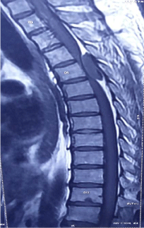
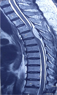
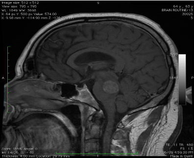
vertebra. Thoracic spine MRI revealed 34.3 × 27.5 × 10.5 mm, well-defined extradural mass at D5
body level, iso-intense to spinal cord on T1 and hyper-intense on T2-weighted images. Spinal cord
at the level of mass was compressed and displaced anteriorly & to the right. Mass extended into
neural foramina at the D5-D6 level. Nerve roots at this level were not definable separately from
the mass (Figure 1,2,3 and 4). Patient underwent D5-D6 laminectomy. Tumor was bluish pink and
was moderately vascular with well defined margin .It was firm in consistency and was purely in
extradural space .The tumor extended along the D5 nerve root on left side. It was excised en-bloc.
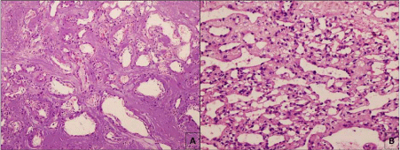
The histopathological examination of excised specimen revealed fibrocollagenous and
fibroadipose tissue. The tissue also showed a dilated irregular vascular channel lined by flattened
epithelium separated by stroma. These features were consistent with capillary hemangioma (Figure 5).
Postoperatively, significant improvement in power of both lower limbs was observed in the
patient along with disappearance of back pain.
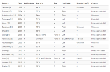
Table 1
Figure 1
Figure 2
Figure 3
Figure 3
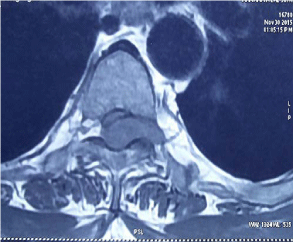
Mri axial section t1 image showing isointense mass extending into
left neural foramino, cord is also compressed anteriorly & towards right.
Discussion
Spinal cord tumors comprise about 15% of all central nervous
system (CNS) neoplasm. Spinal vascular tumors may be classified as capillary telangiectasias, cavernous angioma, capillary hemangiomas,
arteriovenous malformations or venous malformations [3]. The
neuro epithelium, ontogenetically giving rise to the distal spinal
cord, is of mesodermal origin. Frequently, tumors at the level of the
conus medullaris and cauda equina contain mesodermal elements.
The occurrence of vascular lesions involving the conus medullaris
and cauda roots in a metameric distribution has occasionally been
recognized. Capillary hemangioma, one of the spinal vascular tumors,
is characterized by a lobular architecture, with each lobule separated
by septa of fibrous connective tissue and consisting of a myriad of
small and very small capillaries lined by endothelial cells [4]. Because
it is usually well demarcated from the surrounding parenchyma by a connective tissue capsule and reveals mild to moderate mitotic
activity, capillary hemangioma can be classified into a benign vascular
tumor or tumor-like lesion, despite the lack of precise understanding
of the details of its development and growth [4]. Spinal epidural
hemangiomas account for 4% of all spinal epidural tumors, mostly
occurring as a primary lesion in the vertebral bone [5]. Though spinal
epidural hemangioma itself is a very rare variety of tumor the capillary
variety is far rarer than cavernous type, according to literature there
are 80 reported cases of cavernous epidural hemangioma while on the
contrary only 9 cases of capillary hemangioma reported in literature
[1,6].
Patient with epidural hemangioma can present with slow and
progressive spinal cord syndrome (most common presentation)
Patients can also present with acute spinal cord syndrome (due to
acute hemorrhage), backache and radiculopathy [7]. There is one case
report in which patient presented with lumber disc herniation [8].
Our patient presented with back pain & radiculopathy. Patients suffer
from progressive myelopathy and early treatment will prevent any
residual neurological deficits [9]. Complete en-bloc resection is the
treatment of choice [9].
Conclusion
Epidural hemangioma is very rare but it should be included in differential diagnosis of epidural mass. Since these are benign lesions so en-bloc excision cures the patient.
Figure 4
Figure 4
Mri axial section t2 image showing hyperintense mass extending
into left neural foramino at d5-6 level.
Figure 5
Figure 5
The photomicrographs showing histological features of the
case (a) multiple thin walled dilated vascular channels separated by thickened
dura, some of which shows rbcs within the lumen (hex100) (b) conglomerate
of the variable sized vascular channels lined by endothelial layer, (hex100)
consistent with capillary hemangioma.
References
- Egu K, Kinata-Bambino S, Mounadi M, Rachid El Maaqili M, El Abbadi N. Lombosacral epidural capillary hemangioma mimicking a dumbbell-shaped neurinoma: A case report and review of the literature. Neurochirugie. 2016; 62: 113-117.
- Choi BY, Chang KH, Choe G, Han MH, Park SW, Yu IK, et al. Spinal intradural extramedullary capillary hemangioma: MR imaging findings. AJNR Am J Neuroradiol. 2001; 22: 799-802.
- Russel DS, Rubinstein LJ. Pathology of tumors of the nervous system. Ed 5. Baltimore: William & Wilkins; 1989. pp. 730–736.
- Enzinger FM, Weiss SW. Benign tumors and tumorlike lesions of blood vessels. In: Enzinger FM, Weiss SW (eds) Soft tissue tumors, 3rd edn. St Louis, Mosby, 1995. pp 579–626.
- Aoyagi N, Kojima K, Kasai H. Review of Spinal Epidural Cavernous Hemangioma. Neurologia medico-chirurgica. 2003; 43: 471-476.
- Rahman A, Hoque SU, Bhandari PB, Abu Obaida AS. Spinal extradural cavernous haemangioma in an elderly man. BMJ Case Rep. 2012.
- Antunes A, Beck MF, Strapasson AC, Franciscatto AC, Franzoi M. Extradural cavernous hemangioma of thoracic spine. Arq Neuropsiquiatr. 2011; 69: 720-721.
- Tekin T, Bayrakli F, Simsek H, Colak A, Kutlay M, Demircan MN. Lumbar epidural capillary hemangioma presenting as lumbar disc herniation disease: case report. Spine (Phila Pa 1976). 2008; 33: E795-E797.
- García-Pallero MA, Torres CV, García-Navarrete E, Gordillo C, Delgado J, Penanes JR, et al. Dumbbell-Shaped Epidural Capillary Hemangioma Presenting as a Lung Mass: Case Report and Review of the Literature. Spine (Phila Pa 1976). 2015; 40: E849-E853.

