Case Report
Plasma Cell Type Localized Intraorbital Castleman's Disease: A Case Report
He Jie1
1Department of Radiology, Hebei Medical University, China
2Department of Pathology, Hebei Medical University, China
*Corresponding author: He Jie, Department of Radiology, The Third Hospital of Hebei Medical University No.139, ziqiang road, Shijiazhuang, Hebei Province, PR. China
Published: 24 Aug 2016
Cite this article as: Jie H, Shiling L, Yang D. Plasma Cell
Type Localized Intraorbital Castleman's
Disease: A Case Report. Ann Clin Case
Rep. 2016; 1: 1096.
Abstract
A 76-year-old male presented with a 3-month history of progressive mass in the right eye area. Excisional biopsy revealed a well-circumscribed mass, histopathological and immunohistochemical
studies of the orbital mass reveal the features consistent with plasma cell type Castleman's disease
(CD). Plasma cell type is not commonly found than hyaline-vascular type in the two types of CD.
In the orbit, CD is extremely rare with few reported cases. We report this patient with localized
intraorbital Castleman's disease.
Keywords: Castleman's disease; Orbit; Magnetic resonance imaging
Introduction
Castleman’s disease (CD) is a rare atypical lymphoproliferative disorder. It was first reported by Benjamin Castleman in 1954 [1]. CD usually presents in the mediastinum (60-75%). In the orbit, CD is extremely rare with few reported cases [2-5]. We report this patient with localized intraorbital Castleman's disease.
Case Presentation
A 76-year-old Chinese male presented with a 3-month history of progressive mass in the right
eye area. Ophthalmic examination revealed a soft tissue mass, which was well- circumscribed and
unmovable in the inferior eyelid.
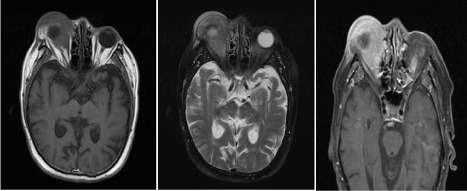
Magnetic resonance imaging (MRI) demonstrated a soft tissue mass in the right orbit located in
the posterior and the lower aspect of the eyeball. MRI presented T1 and T2 isointensity and slightly
hyperintensity in fat suppressed sequence. The mass which grew around the eyeball, extruded beyond
the orbit and involved in the internal and external pyramidal is muscle. The eyeball was compressed
and dislocated. There was obvious and homogeneous enhancement (Figure 1). The MRI diagnosis
suggested that the benign lesion located in the orbit maybe inflammatory pseudotumor.
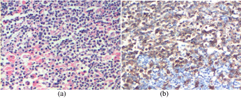
Pathological examination: Microscopically, the soft tissue mass were found to be irregular and
grey-red in color. Part of the tissue showed node-like structures. Characteristically, the interfollicular
zones showed numerous plasma cells and Russell bodies. Additionally, immunohistochemical
staining revealed that the majority of the plasma cells expressed CD38 positive stain (Figure 2).
Histopathological and immunohistochemical studies of the orbital mass reveal the features
consistent with plasma cell type CD.
Figure 1
Figure 1
MRI axial scan (A: T1WI; B: T2WI; C: T1 contrast enhancement) showing an extraconal soft tissue mass
with obvious and homogeneous enhancement in the right orbit.
Figure 2
Figure 2
Microscopic view of the mass showing hyperplasma of solid,
confluent sheets of plasma cell and Russel bodies in the interfollicular zones
(HE staining 4×100). B: CD38 staining showing positive stain (20×10).
Discussion
Clinically, CD occurs in localized and multicentric forms.
Pathologically, it is subdivided into two forms: hyaline-vascular type
and plasma cell type. Hyaline-vascular type is most commonly found
(90%). The characteristic histopathological features of this form are
the presence of abnormal follicles with atrophic hyalinized follicular
centre and a broad mantle zone of small lymphocytes in a concentric
or onion skin arrangement.
The characteristic histopathological feature of the plasma cell
type form is the presence of solid, confluent sheets of plasma cells
in the interfollicular areas. The follicular centres are usually enlarged
and hyperplastic.
The unique sign of this case is the intraorbital soft mass. The
differential diagnosis with other orbital mass consist of: 1. Diffuse
inflammatory pseudotumor: The mass presents medium andhypointensity in T1 weighted image and medium and hyperintensity
in T2 weighted image, which shows the medium enhancement and
presents thickening of optic nerves and swelling of lacrimal gland.
2. Cavernous hemangioma. The mass presents hypointensity or
isointensity in T1 weighted image and hyperintensity in T2 weighted
image, which shows the character of gradual enhancement.
The case reported in this paper presented isointensity in T1 and T2
weighted image, which was different from the other mass in the orbit.
The enhancement was different from cavernous hemangioma. Optic
nerve and cellulitis gland were not involved, which was different
from inflammatory pseudotumor. It is therefore suggested that CD
should be considered when the atypical mass was found in the orbit.
Histopathological examination needs to be conducted in order to
confirm the diagnosis.
References
- Castleman B, Towne VW. Case records of the Massachusetts General Hospital: Case No. 40231. N Engl J Med. 1954; 250: 1001-1005.
- Park KS, Choi YJ, Song KS. Hyaline-vascular type Castleman's disease involving both orbits. Acta Ophthalmol Scand. 2002; 80: 537-539.
- Kim U, Hwang JM. Optic neuropathy associated with Castleman disease. Korean J Ophthalmol. 2010; 24: 256-259.
- Jáñez L, Taban M, Wong CA, Ranganath K, Douglas RS, Goldberg RA. Localized Intraorbital Castleman's disease: a case report. Orbit. 2010; 29: 158-160.
- Venizelos I, Papathomas TG, Papathanasiou M, Cheva A, Garypidou V, Coupland S. Orbital involvement in Castleman disease. Surv Ophthalmol. 2010; 55: 247-255.


