Case Report
Citrobacter Meningitis/Cerebritis: A Case Report
Kurian ST*, Halpin J and Chin Brian M
Diagnostic Radiology Residency Program, University of Missouri Kansas City, USA
*Corresponding author: Sarah T. Kurian, University of Missouri Kansas City, Diagnostic Radiology Residency Program c/o Julie McCollum, 4401 Wornall Road, Kansas City, MO 64111, USA
Published: 24 Aug 2016
Cite this article as: Kurian ST, Halpin J, Chin Brian M.
Citrobacter Meningitis/Cerebritis: A
Case Report. Ann Clin Case Rep. 2016;
1: 1093.
Abstract
Citrobacter koseri (CK) is a facultative anaerobe that causes an uncommon, yet potentially
neurologically devastating, form of neonatal meningitis/cerebritis. On imaging it often forms
a characteristic polygonal-shaped abscess. A high degree of suspicion is necessary to make the
diagnosis. One such case of a neonate is presented, illustrating the key imaging features and
importance of serial neuroimaging.
Keywords: Citrobacter koseri; Polygonal abscess; Intra-axial abscess; Meningitis; Cerebritis
Introduction
Citrobacter koseri (CK) is a facultative anaerobe that causes an uncommon, yet potentially neurologically devastating, form of neonatal meningitis/cerebritis. On imaging it often forms a characteristic polygonal-shaped abscess . A high degree of suspicion is necessary to make the diagnosis. One such case of a neonate is presented, illustrating the key imaging features and importance of serial neuroimaging.
Case Presentation
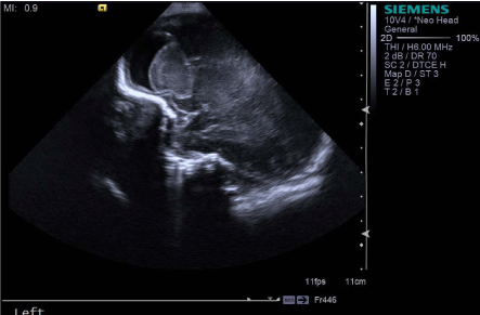
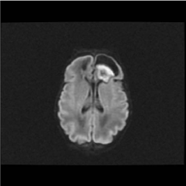
Born at 28 weeks, the patient underwent a septic workup for leukocytosis on day of life 7, without a source of infection found, including CSF analysis. The leukocytosis persisted despite 3 weeks of broad antibiotic therapy. A head ultrasound obtained at day of life 36 showed complex bifrontal heterogeneous intra-axial masses/collections. Noncontrast MRI showed imaging features consistent with cerebritis with cortical/sub-cortical white matter edema and a polygonal-shaped intracranial abscess in the left frontal region. On serial imaging, there was continued growth of the intra-axial collections, so the patient was transferred to a children's hospital for continued care.
Discussion
As demonstrated in this case, serial neuroimaging in the setting of persistent leukocytosis is important even with negative CSF studies. Such imaging can detect complications of meningitis/ cerebritis, such as extra-axial collections, intra-axial abscesses, midline shift, hydrocephalus, and even pneumocephalus . While CSF may be negative, CK meningitis/cerebritis should be suspected if the characteristic polygonal abscess formation is seen. Early imaging findings of cerebritis include increased echogenicity of the subcortical periventricular white matter on head ultrasound and a complex appearing, intra-axial mass as cerebritis progresses and an abscessdevelops. On MRI, T2/FLAIR hyperintense signal is present in the cortical gray and subcortical/deep white matter in cerebritis, and diffusion restriction, upon development of an intra-axial abscess. If contrast is administered, rim enhancement is seen with an abscess.
Figure 1
Figure 1
Head Ultrasound: Hyperechoic intra-axial collection/mass is seen in the left frontal region.
Figure 2
Figure 2
MRI Axial DWI (diffusion): Left frontal lobe, polygonal shaped
abscess with central area of diffusion restriction.
Conclusion
Familiarity with Citrobacter meningitis/cerebritis and its complications is important, as there is a high mortality rate and risk of long-term CNS damage in neonates. Even if CSF fluid does not grow the organism, it should be suspected if the characteristic appearance is seen. Antibiotics are the mainstay of treatment. If medical management is inadequate for treatment, other treatment options include abscess drainage and intraventricular urokinase for loculated hydrocephalus .
References
- Barkovich A. Diagnostic Imaging: Pediatric Neuroradiology. 2nd ed. Salt Lake City: Amirsys; 2014.
- Alviedo Joseph N, Beena G. Sood, Jacob V. Aranda, Cristie Becker. Diffuse Pneumocephalus in Neonatal Citrobacter Meningitis. Pediatrics. 2006; 118: e1576-e1579.
- Martínez-Lage Juan F, Laura Martínez-Lage Azorín, María José Almagro, María Encarnación Bastida, Susana Reyes, Cinthia Tellez. Citrobacter Koseri Meningitis: A Neurosurgical Condition?. Eur J Paediatr Neurol. 2010; 14: 360-63.


