Case Report
Herpes Zoster Ophthalmicus with Isolated Paralytic Mydriasis
Casal IA1*, Monteiro S1, Borges T1, Vale C1, Friande A1, Araújo M1 and Beirão JM1,2
1Centro Hospitalar do Porto – Hospital de Santo António, Portugal
2Instituto de Ciências Biomédicas Abel Salazar, Universidade Porto, Portugal
*Corresponding author: Inês Casal, Department of Ophtalmology, Porto Hospital Centre, Portugal Rua Prof. Bento Jesus Caraça, Nº189, Hab. 65, 4200-131 Porto, Portugal
Published: 16 Aug, 2016
Cite this article as: Casal IA, Monteiro S, Borges T, Vale
C, Friande A, Araújo M, et al. Herpes
Zoster Ophthalmicus with Isolated
Paralytic Mydriasis. Ann Clin Case Rep.
2016; 1: 1082.
Abstract
Introduction: Herpes zoster ophthalmicus occurs when there is a reactivation of herpes 3 lying latent
in the ophthalmic division of the trigeminal nerve. We report a case of an 86 year-old female patient
with an orbital cellulitis with post-septal involvement admitted in our departement for topical and
intravenous antibiotics. On the fourth day of admission vesicular lesions began to emerge in the
left side of the face and scalp not exceeding the mid line and treatment for herpes zoster withoral
acyclovir and topic ganciclovir was initiated. Then an anisocoria was noticed and in the left pupil
the direct and consensual reflexes were abolished as well as the accommodation reflex. There was
no intraocular hypertension, anterioror posterior synechiae; no external ophthalmoplegia, ptosisor
diplopia. The brain CT revealed no relevant alterations. The patient was discharged after eighteen
days of hospitalization, maintaining the paralytic mydriasis of the left pupil.
Discussion: Isolated paralytic mydriasis as the only complication after herpes zoster ophthalmicusis
extremely rare. The responsible mechanisms are notfully understood. It is thought that the cause
is, among other factors, the involvement of the pupillary fibers for light and accommodationconvergence,
with no damage of the motor fibers.
Introduction
Herpes zoster ophthalmicus occurs when there is a reactivation of herpes 3 (varicella-zoster) lying latent in the ophthalmic division of the trigeminal nerve, and this involvement occurs in about 10-25% of the cases [1]. Half of these will have ocular involvement [2,3], being the symptoms varied and caused by the perineural and intraneural inflammation of sensory nerves [4]. The various manifestations obey the midline and depend on the branches of the trigeminal nerve that are affected (supraorbital, lacrimal or nasociliary). The nasociliary branch innervates the globe, so when it is affected there is a greater probability of serious ocular involvement and complications. The classical sign of these involvement is the “Hutchinson's sign” – involvement of the tip of the nose - and these patients have twice the incidence of ocular affection [5]. Ocular involvement may be at the eyelids with a papulo-vesicular rash; blepharoconjunctivitis; episcleritis/scleritis; corneal involvement with dendritic keratitis, stromal and neurotrophic keratitis; uveitis; intraocular hypertension and even acute retinal necrosis [6]. The cranial nerves involvement has been reported as optic neuritis, total external or internal ophthalmoplegias, and isolated third, fourth and sixth cranial nerve palsies; rarely associated with pupillary involvement [6]. The extraocular muscle palsies occur in 7 to 31% of the patients: the third nerve is the most commonly affected, followed by the sixth nerve and the fourth is the least affected [7-9]. Aging, nutrition deficits, immune compromised status and other factors such as fatigue and stress (physical / emotional) may precipitate an episode of herpes zoster. Some of the serious and permanent sequelae of herpes zoster ophthalmicus are debilitating pain, chronic ocular inflammation or even loss of vision [10].
Case Presentation
We present a case of an 86 year-old female patient that went to our Emergency department
with eyelid edema associated with conjunctival hyperemia in the left eye with 3 days of evolution.
Physical examination showed mild erythema and edema of the left cheek and more marked and
painful eyelid edema. Slit-lamp examination showed mild conjunctival hyperemia and chemosis
with mucopurulent secretions in the lower lashes, pseudophakia and no signs of anterior chamber
inflammation or other alterations. The pupils were regular, direct and consensual light reflexes
were preserved, she had no proptosis, limitation of ocular movements or diplopia. The visual acuity of the left eye was 0.3 (decimal scale) and the right eye 0.8, without any correction. The computed tomography (CT) scan of the orbits revealed orbital cellulitis with post-septal involvement. The patient was admitted for treatment with topical of loxacin and intravenous antibiotic therapy with vancomycin 1 g bid and ceftriaxone 1 g bid. On the fourth day post admission, began to emerge vesicular lesions in the left side of the face and scalp that did not exceed the midline, being very suggestive of herpes zoster ophthalmicus. The patient presented with lesions in the tip of the nose - Hutchinson's sign - suggesting involvement of the nasociliary branch and greater probability of more serious ocular involvement. The remaining examination just showed mild superficial keratitis, without signs of anterior chamber inflammation, ocular hypertension or posterior segment alterations; and we began therapy with oral acyclovir 800 mg, 5 times per day and ocular topic ganciclovir q4h. On the sixth day post admission the skin lesions were improving and at different stages of evolution, there was an improvement of the face and eyelid edema, but the left pupil was almost not reactive to light, still with no signs of anterior chamber inflammation or ocular hypertension. Oral corticosteroid therapy was initiated. On the ninth day of hospitalization and despite symptomatic improvement, we objectified an anisocoria (left pupil 3 mm bigger than the right) which decreased in scotopic conditions, with the left pupil almost with no light reaction (Figure 1). Slit-lamp examination showed a mild conjunctival hyperemia and superficial keratitis in the lower half of the cornea; there were no alterations of the iris and no signs of anterior chamber inflammation, anterior or posterior synechiae. The intraocular pressure was 15/16 mmHg and the visual acuityofthelefteyedecreased to 0.1.Brain CT scan revealed ischemic leukoencephalopathy and enlargement of the cerebrospinal fluid circulation pathways by atrophy - changes consistent with the age of the patient. The neurology examination showed preserved higher functions; algic hypoesthesia in the territory of the left first division of the trigeminal nerve and anisocoria described as above: both pupils were regular, and the right pupil had direct and consensual light reflexes preserved but in the left pupil, the direct and consensual light reflexes were abolished as well as the accommodation reflex. There were no external ophthalmoplegia, ptosisor diplopia (Figure 2).
The patient was discharged after eighteen days of hospitalization, asymptomatic, with almost complete resolution of the skin and scalp lesions and no eyelid edema. She completed 13 days of intravenous therapy with vancomycin and ceftriaxone; 14 days of oral acyclovir and topical ganciclovir and 11 days of oral corticosteroid therapy. Currently, six months after discharge, the patient has a visual acuity of the left eye of 0.3; the slit lamp examination shows only a slight posterior capsule opacification and maintains the anisocoria with paralytic mydriasis of the left pupil.
Figure 1
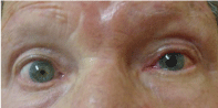
Figure 1
Anisocoria with the left pupil bigger than the right.
Figure 2
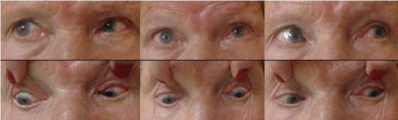
Figure 2
No external ophthalmoplegiaorptosis.
Discussion
Isolated paralytic mydriasis as the only complication after herpes zoster ophthalmicus is extremely rare, with only few cases reported. Usually pupillary paralysisis reported associated with some degree of ophthalmoplegias (total, external/internal) or isolated palsies of the third, fourth or sixth nerves [2]. The responsible mechanisms are still unclear. In this case we excluded some of the inflammatory causes as inflammation of the anterior chamber with iritis, iris atrophy, anterior and posterior synechiae; as well as ocular hypertension.Various the ories have been postulated–Denny-Brown et al. [11] described that motor neuritis was independent of the inflammation of any ganglion; Kreibig [12] concluded that perivasculitis-myositis was responsible for the extraocular palsies rather than a neural cause; Godtfredsen [13] postulated that the involvement of the cranial nerves was due to contiguous intracavernous radiculo meningitis; and the major cause could also be ischaemia due to occlusive vasculitis [2]. The cause of the isolated paralytic mydriasis is probably a combination of these mechanisms (separately or simultaneously) that led to aparcial third nerve palsy with spared motor fibers but with the involvement of the pupillary fibers for light and accommodation-convergence [2]. These paralytic lesions tend to be temporary and resolve spontaneously; and there is no specific treatment described in the literature.
References
- Ragozzino MW, Melton LJ 3d, Kurland LT, Chu CP, Perry HO. Population-based study of herpes zoster and its sequelae. Medicine. 1982; 61: 310–316.
- Czyz CN, Bacon TS, Petrie TP, Justice JD, Cahill KV. Isolated, complete paralytic mydriasis secondary to herpes zoster ophthalmicus. Pract Neurol 2013; 13:183-184.
- Marsh PJ, Dulley B, Kelly V. External ocular motor palsies in ophthalmic zoster: a review, Br J Ophthalmol. 1977; 61: 677-682.
- Naumann G, Gass JD, Font RL. Histopathology of herpes zoster ophthalmicus. Am J Ophthalmol. 1968; 65: 533–541.
- Harding SP, Lipton JR, Wells JC. Natural history of herpes zoster ophthalmicus: predictors of postherpetic neuralgia and ocular involvement. Br J Ophthalmol. 1987; 71: 353–358.
- Kanski JJ. Herpes zoster ophthalmicus. In: Kanski JJ, Nischal KK, Milewski SA, eds. Ophthalmology: clinical signs and differential diagnosis. Philadelphia: Mosby, 1999.
- Hunt JR. The paralytic complications of herpes zoster of the cephalic extremity. JAMA. 1909; 53: 1456–1460.
- Edgerton AE. Herpes zoster ophthalmicus: report of cases and review of literature. Trans Am Ophthalmol Soc. 1942; 40: 390–439.
- Kelly V, Dulley B. Ocular motor defects associated with herpes zoster ophthalmicus. In: Moore S, Mein J, Stockbridge L, editors. Orthopotics-Past, Present and Future. Symposium Specialist. New York: Stratton Intercontinental Medical Book Corp. 1976; 367–377.
- Shaikh S, Christopher N. Evaluation and Management of Herpes Zoster Ophthalmicus. Am Fam Physician. 2002; 66: 1723-1730.
- Denny-Brown D, Adams RD, Fitzgerald PJ. Pathologic features of herpes zoster: a note on "genicular herpes". Arch Neurol Psychiatry. 1944; 51: 216–231.
- Kreibig W. Die Zosterekrankung des Auges. Klin Monatsbl Augenheikd. 1959; 135: 1–31.
- Godtfredsen E. Pathogenesis of cranial nerve lesions, notably ophthalmoplegias, complicating herpes zoster opthalmicus. Acta Psychiatr Neurol. 1948; 23: 69–77.