Case Report
Multiple Contiguous Avulsion Fractures of Thoracic Spinous Processes: A Clay Shoveler’s Variant
Ali Bakhsh* and Himanshu Sharma
Department of Orthopedics, The Peninsula College of Medicine and Dentistry, UK
*Corresponding author: Ali Bakhsh, Department of Orthopedics, The Peninsula College of Medicine and Dentistry, John Bull Building, Plymouth, PL6 8BU, UK
Published: 08 Aug, 2016
Cite this article as: Bakhsh A, Sharma H. Multiple
Contiguous Avulsion Fractures of
Thoracic Spinous Processes: A Clay
Shoveler’s Variant. Ann Clin Case Rep.
2016; 1: 1076.
Abstract
Avulsion fracture of the spinous process, also known as a clay-shoveler’s fracture, is usually seen at a single level or dual adjacent spinal levels in the lower cervical or cervicothoracic junction. We report a 49-year old lady presented late with multiple contiguous clay-shoveler’s fracture between T4 and T8 on MRI scan with negative initial plain radiography. Thoracic spine x-rays might miss the avulsion fractures. With persistent pain and crepitus in the cervicothoracic spine following sudden forced hyper-flexion & rotational trauma, further imaging in the form of MRI scan with or without CT scan is recommended. Disproportionate pain in the initial post-injury period should raise a high index of suspicion. Prolonged recovery period of 6-9 months should be provided to the patients for preferred conservative treatment.
Keywords: Spinous process; Fracture; Avulsion
Introduction
A clay-shoveler’s fracture is an avulsion facture of the spinous process classically seen at C6 or
C7 level [1]. It is most commonly associated with manual labour. Clay-shoveler’s fracture may occur
through direct trauma or by shear forces. The most common cause is hyperflexion and rotation of
the spine [2]. The mechanism of injury is believed to be secondary to muscle pull and reflex with
force transmission through the supraspinous ligaments [3] resulting in an avulsion fracture of the
spinous processes.
Multiple and contiguous clay-shoveler’s fractures affecting thoracic spine are exceedingly rare.
We present a 49 year old female who sustained 5 contiguous thoracic avulsion fractures between T4
and T8 vertebrae with accidentally fallen whilst horse-riding. As far as we are aware, we believe that
our case is the fifth case reported in literature of an isolated spinous process fracture involving four
or more levels in the thoracic vertebrae.
The author has obtained the patient’s informed written consent for print and electronic
publication of the case report.
Case Presentation
A 49 year old lady presented with constant inter-scapular back pain, worse when working
overhead and on bending or twisting her thoracic spine. The patient rated her pain 9/10 on the
Visual Analogue Scale (VAS). There were no red flags and bladder and bowel functions were
normal. She reported a horse-riding accident 4weeks previously where she fell onto her left side
from a height of 6 feet. She experienced instantaneous pain at the time between her shoulder blades
with instantaneous crepitus. She confirmed that the pain was 10/10 on the VAS immediately after
the incident. The patient described right arm tingling and numbness affecting the middle, ring and
little fingers on her right side persisting consistently since her accident.
The patient attended the Emergency Department the same day of her fall where she was
examined and plain X-ray of cervical and thoracic spine were taken. The X-rays did not show any
bony injuries (Figure 1). She was prescribed opiate and non-steroidal anti-inflammatory analgesia
and self-medicated with amitriptyline. The pain persisted and 1 week later the patient attended her
General Practitioner who prescribed Paracetamol, Co-codamol and Naproxen. 2 weeks after her fall,
the patient self-referred to physiotherapy who declined treatment due to presentation of her severe
symptoms and the crepitus in her thoracic spine. A MRI scan was requested by the physiotherapist.
At this point, the patient described her pain was unmanageable with simple analgesia and she
required time off work. Past medical history is insignificant.
On examination, there was localised tenderness between her shoulder blades right from the T4 to the T8 area with some crepitus felt. Thoracic spine rotations were painful beyond 50%. There was altered sensation in the paraspinal muscle area with some dull sensation. Bilateral upper limb neurological examination confirmed presence of paraesthesia on the right side in the ulnar nerve distribution. The range of movement in the cervical spine was limited in the terminal range along with some crepitus.
MRI scan of the cervical spine (Figure 2) confirmed multi-level degenerative changes and presence of multi-level foraminal stenosis but no obvious nerve root compression as such. MRI scan of the thoracic spine (Figure 3A and B) confirmed presence of avulsion fractures of the spinal processes of the T4, T5, T6, T7 and T8 levels. Spinal column integrity was intact and there was no injury to the spinal column or nerve roots was noted.
This patient was managed conservatively. We advised her to wear a thoraco-lumbo-sacral orthosis (TSLO) brace for 4 weeks aiming to gradually wean-off after a further 4 weeks. During the weaning period, she should start gentle physiotherapy with a view to gradually mobilise a range of motions. Thereafter, she should gradually work to build up her core muscles.
Figure 1
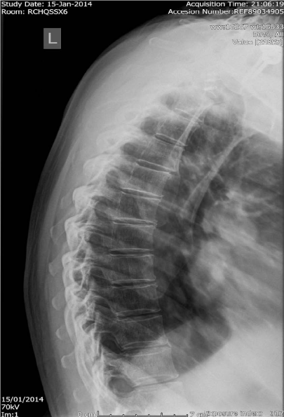
Figure 1
Lateral thoracic spine x-ray shows no fracture.
Figure 2
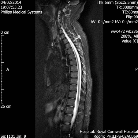
Figure 2
Lateral MRI scan of whole spine showing multi-level degenerative changes and presence of multi-level foraminal stenosis.
Figure 3
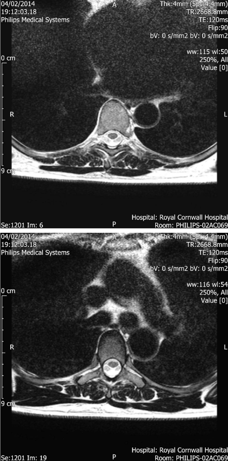
Figure 3a, 3b
Axial MRI scan showing discontinuity and mild displacement to the right of the midline of the tips of the spinous processes of T4 (3a), T5 and T6 and T7 (3b). This can only be seen on the axial views and is in keeping with spinous process fractures. There is also oedema of the spinous process of T8, suspicious of a fracture, but this level is not included on the axial images.
Discussion
MRI studies have a role in diagnosis of such patients. Immobility and rehabilitation continue to be the mainstay of management. The clinical presentation consists mainly of tenderness and pain in the cervical and thoracic spine. Usually, there are no neurological deficits detected.
Traditionally, plain radiography has been adequate to rule out spinous fractures. However, as with our patient, avulsion fractures can be difficult to assess on plain radiographs. There is a definite role for MRI studies [4] in patients with severe pain but no abnormalities detected on plain radiography.
CT scan could be of use for depicting detailed bony anatomy, however, as these are avulsion fractures, haematoma in the paraspinal muscles and ligamentous disruption could be identified much better with MRI scan though. In addition, this lady had upper limb neurological manifestations post-injury as well.
These injuries are known to be painful but stable. Generally, these types of fractures are treated conservatively. Surgical intervention is not advisable and not likely to change the outcome of patient’s long-term residual symptoms [5]. Immobilization with a thoraco-lumbo-sacral-orthosis and restriction of physical activity for 4 to 6 weeks is recommended. Thereafter, gentle physiotherapy is recommended with a view to gradual mobilisation. Generally there is a good outcome however symptoms of aches and stiffness between the shoulder blades may persist.
Our case highlights important messages. Firstly, clay-shoveler fractures may produce severe pain which is out of proportion to clinical signs. There needs to be a high-index of suspicion for a clay-shoveler’s fractures in shear injuries and a lower threshold for MRI studies.
Correct diagnosis is essential so a comprehensive rehabilitation plan can be offered. The mainstay of management is initial immobilisation for 4 weeks with rehabilitation by way of gentle physiotherapy. The majority of patients treated conservatively have good clinical outcomes with minimal residual symptoms [6].
References
- Akhaddar A, El-asri A, Boucetta M. Multiple isolated thoracic spinous process fractures (Clay-Shoveler's fracture) Spine J. 2011; 11: 458–459.
- Kim SY, Chung SK, Kim DY. Multiple cervical spinous process fractures in a novice golf player. J Korean Neurosurg Soc. 2012; 52: 570-573.
- Kose KC. Case report: The impact of pseudoarthrosis on clinical outcome in isolated spinous process fractures of six adjacent level thoracic vertebrae. Med Gen Med 2006; 8: 67.
- Solaroğlu I, Kaptanoğlu E, Okutan O, Besknnakli E. Multiple isolated spinous process fracture (Clay-shoveler's fracture) of cervical spine: a case report. Ulus TravmaAcilCerrahiDerg. 2007; 13: 162–164.
- Kaloostian PE, Kim JE, Calabresi PA, Bydon A, Witham T. Clay-shoveler's fracture during indoor rock climbing. Orthopedics 2013; 36: e381-383.
- Han SR, Sohn MJ. Twelve contiguous spinous process fracture of cervico-thoracic spine. Korean J Spine 2014; 11: 212-213.