Case Report
Liver Metastasis from a Renal Epithelioid Angiomyolipoma: A Case Report
Akbar A1, Mushtaq S2, Syed AA1 and Hanif F1*
1Department of Surgical Oncology, Shaukat Khanum Memorial Cancer Hospital & Research Center, Pakistan
2Department of Pathology, Shaukat Khanum Memorial Cancer Hospital & Research Center, Pakistan
*Corresponding author: Faisal Hanif, Department of Surgical Oncology, Shaukat Khanum Memorial Cancer Hospital and Research Centre, Johar Town, Lahore, Pakistan
Published: 08 Aug, 2016
Cite this article as: Akbar A, Mushtaq S, Syed AA, Hanif
F. Liver Metastasis from a Renal
Epithelioid Angiomyolipoma: A Case
Report. Ann Clin Case Rep. 2016; 1:
1074.
Abstract
Epithelioid angiomyolipoma (EAML) are rare tumours and belong to a family of tumours known as perivascular epitheloid cell tumors which also include classic angiomyolipoma. Renal EAML have a female preponderance and are often difficult to diagnose as they mimic renal cell carcinoma due to similar clinical symptoms and imaging. There are few anecdotal reports on liver metastasis from renal EAML highlighting the fact that the patients should be followed up with imaging so that any metastasis can be picked up in time and treated on its merit. We present a case report of a 20 year old male who developed liver metastasis needing resection, 3 years after left nephrectomy for an EAML.
Keywords: Angiomyolipma; Renal epithelioid angiomyolipoma; Metastasis; Liver metastasis
Introduction
Epithelioid angiomyolipoma (EAML) represents rare family of tumors known as PECOmas (perivascular epitheloid cell tumors) which also include classic angiomyolipoma. EAML are often difficult to diagnose as they mimic Renal Cell Carcinoma due to similar clinical symptoms and imaging [1]. There are few anecdotal reports on liver metastasis from renal EAML highlighting the fact that the patients should be followed up with imaging so that any metastasis can be picked up in time and treated on its merit. Here we report a case of EAML of kidney with liver metastasis.
Case Presentation
A 20 year old male presented with pain in left lumbar region for 6 weeks. His abdominal
examination was unremarkable but his CT scan abdomen showed left renal tumor involving upper
& middle poles with areas of calcification, extending beyond renal fascia, abutting left flank with no
evidence of renal vein or inferior vena cava thrombosis, no abdominal or pelvic lymphadenopathy.
Left radical nephrectomy was carried out and on histopathology a diagnosis of EAML was confirmed.
He was followed up with yearly imaging with no evidence of recurrence for 3 years. CT scan at
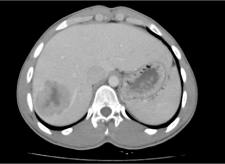
4 years showed a lobulated primarily solid hypodense mass with central necrosis in segment VI and
VII of liver having maximum dimension of 57 mm (Figure 1).
The patient underwent ultrasound guided fine needle aspiration cytology of the liver lesion
which was consistent with metastatic Epitheliod Angiomylipoma, Decision in the multidisciplinary
team meeting was to rule out tuberous sclerosis (TS) and perform liver resection of the metastatic
deposit. MRI brain was performed which was negative for any features of TS. He underwent liver
resection to remove the tumor. Histopathology confirmed 6.0 cm x 5.5 cm x 4.6cm metastatic EAML
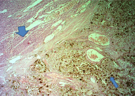
with clear resection margins. Microscopic evaluation revealed cellular neoplasm with proliferation
of epithelioid cells having abundant granular and pigmented cytoplasm. Tumor cells were arranged
in sheets and numerous dilated vessels with perivascular cuffing was present. Individual cells were
polygonal with vesicular nuclei and prominent nucleoli. There were multiple areas with clear cell
morphology and mitotic figures were present with strongly positive HMB45 (Figure 2). He has been
doing well 6 months after surgery with no evidence of recurrence in follow-up imaging.
Figure 1
Figure 2
Figure 2
Broad arrow indicating normal liver, thin arrow showing a circumscribed neoplasm with abundant hemosiderin pigment, round to oval cells with clear to vaculated cytoplasm. Numerous dilated vessels present.
Discussion
EAML is a subtype of angiomyolipma which is characterized by epitheloid cell proliferation. It is
most commonly sporadic, however association with TS has been reported in 22% of cases [2]. These
tumors are of mesenchymal origin with typical perivascular epithelioid cell differentiation. EAML
show female predominance with a ratio of 6.5:1 [3].
Patients in EAML are younger as compared to AML (38.6 years versus 52.3 years) whereas age of our patient is 20 years. EAML is found to be associated with advanced disease at presentation including extension into the vena cava and right atrium, and metastasis but without lymph node involvement. Nese et al. [2] reported that approximately one half of patients ultimately develop metastatic lesions and one-third of them die because of disease progression. Metastatic sites included liver (63%), lung (25%), peritoneum/mesentery (18.8%), colon (6.3%), pelvis (6.3%), and diaphragm (6.3%) [2].
EAML is difficult to differentiate from RCC clinically and radiologically, it has been described that renal AML is markedly hypoechoic relative to the renal parenchyma. This finding is consistent with presence of fat within the tumor and on CT imaging is thought to be confirmatory for AML. Microscopically, immunohistochemistry can differentiate EAML from RCC, EAML frequently presents with necrosis,
hemorrhage, nuclear atypia, and mitotic activity. Different histological criteria’s are defined to predict malignant potential like large tumor size, presence of necrosis, frequent mitoses, and atypical mitotic figures. Whereas metastasis may be the only definite stigmata of malignancy [4-7].
Liver is the most common reported site of metastasis with EAML followed by lungs [2]. The definitive treatment of EAML is excision of the tumour. No adjuvant treatment has been proven to be effective. Doxorubicin has been tried and newer agents like rapamycin which is mTOR inhibitor have shown promising results [8] but no robust data is available to prove the efficacy of any chemotherapeutic agent. Because of potential for metastasis, patients with renal EAML should be followed up with regular imaging.
Conclusion
All patients with renal EAML should be carefully followed up with radiological imaging after resection of the primary tumour as this tumour can metastasise to other organs including liver.
References
- Svec A, Velenská Z. Renal epithelioid angiomyolipoma--a close mimic of renal cell carcinoma. Report of a case and review of the literature. Pathol Res Pract. 2005; 200: 851-856.
- Nese N, Martignoni G, Fletcher CD, Gupta R, Pan CC, Kim H, et al. Pure epithelioid PEComas (so-called epithelioid angiomyolipoma) of the kidney: A clinicopathologic study of 41 cases: detailed assessment of morphology and risk stratification. Am J Surg Pathol. 2011; 35: 161-176.
- Aydin H, Magi-Galluzzi C, Lane BR, Sercia L, Lopez JI, Rini BI, et al. Renal angiomyolipoma: clinicopathologic study of 194 cases with emphasis on the epithelioid histology and tuberous sclerosis association. Am J Surg Pathol. 2009; 33: 289-297.
- Moudouni SM, Tligui M, Sibony M, Doublet JD, Haab F, Gattegno B, et al. Malignant epithelioid renal angiomyolipoma involving the inferior vena cava in a patient with tuberous sclerosis. Urol Int. 2008; 80: 102-104.
- Bakshi SS, Vishal K, Kalia V, Gill JS. Aggressive renal angiomyolipoma extending into the renal vein and inferior vena cava - an uncommon entity. Br J Radiol. 2011; 84: e166-168.
- Li J, Zhu M, Wang YL. Malignant epithelioid angiomyolipoma of the kidney with pulmonary metastases and p53 gene mutation. World J Surg Oncol. 2012; 10: 213.
- Sato K, Ueda Y, Tachibana H, Miyazawa K, Chikazawa I, Kaji S, et al. Malignant epithelioid angiomyolipoma of the kidney in a patient with tuberous sclerosis: an autopsy case report with p53 gene mutation analysis. Pathol Res Pract. 2008; 204: 771-777.
- Wolff N, Kabbani W, Bradley T, Raj G, Watumull L, Brugarolas J. Sirolimus and temsirolimus for epithelioid angiomyolipoma. J Clin Oncol. 2010; 28: e65-68.


