Case Report
Nasal Cavity and Laryngopharyngeal Erosion Secondary to Intranasal Pain Medication Abuse: A Case Report
Baker A1, Oberman BS1, Warrick J2 and Andrews G1*
1Department of Surgery, The Pennsylvania State University, USA College of Medicine, USA
2Department of Pathology, The Pennsylvania State University, USA
*Corresponding author: Genevieve Andrews, Department of Surgery, Division of Otolaryngology- Head & Neck Surgery, The Pennsylvania State University, College of Medicine, 500 University Drive, H091, Hershey, PA 17033-0850, USA
Published: 05 Aug, 2016
Cite this article as: Baker A, Oberman BS, Warrick
J, Andrews G. Nasal Cavity and
Laryngopharyngeal Erosion Secondary
to Intranasal Pain Medication Abuse: A
Case Report. Ann Clin Case Rep. 2016;
1: 1072.
Abstract
Objectives: Intranasal abuse of prescription medications is well documented; however, few reports
of intranasal acetaminophen abuse exist. We characterize the presentation and manifestations of
intranasal acetaminophen abuse in order to expedite diagnosis and treatment.
Methods: We present a case of a 20-year-old female presented with 6 months of pain, pharyngitis,
hoarseness, and odynophagia secondary to intranasal acetaminophen inhalation, which was initially
treated for sinusitis.
Results: Initial examination showed erosion of nasal, nasopharyngeal, and oropharyngeal structures
resulting from her acetaminophen inhalation. Treatment included debridement of necrotic tissues
and debris, as well as long-term antimicrobials. In follow-up, there was stenosis of supraglottic
structures.
Conclusions: Intranasal acetaminophen abuse can present with signs and symptoms similar to
pharyngitis or sinusitis, with visible crushed pill material. Long-term abuse can lead to the erosion
of upper aerodigestive tract structures.
Introduction
The abuse of prescription pain medicine is prevalent in today’s society. There are approximately
2,500 first-time users of prescription pain medication daily, leading to 2.6 million new patients
prescribed pain medications yearly [1]. Recent data from 2010 suggests 2 million new abusers of
prescription drugs, particularly hydrocodone, each year [2]. According to the National Institute of
Drug Abuse, fifteen percent of high school seniors abused prescription drugs in 2013 [3].
Intranasal abuse of prescription pain medicine is well described. The majority of these patients
present with oronasal mucosal necrosis. During active abuse, patients have characteristic necrosis
with a white, crusty exudate in the nasal cavity often extending into the posterior oropharynx.
Purulent nasal discharge is an additional sign often seen during periods of active abuse. Other
manifestations include perforation of the nasal septum and perforation of the nasal floor or palate.
Non-invasive fungal disease may be seen with diffuse involvement of the nasal mucosa by fungal
elements. This entity is clinically different from the more life-threatening invasive fungal disease
seen in immunocompromised patients [4].
Few reports exist of intranasal abuse of over-the-counter pharmacologic agents such as
acetaminophen. Of particular interest, 24.6 billion doses of acetaminophen were sold in 2008 [5].
We hope to better characterize the presentation and manifestations of this behavior in order to
facilitate earlier diagnosis and initiation of treatment.
Case Report
A 20-year-old female was referred to the outpatient Otolaryngology clinic with complaints of
nasal and throat pain, hoarseness, odynophagia, and headache for six months, with an associated
92-pound weight loss. The patient claimed that her symptoms were related to inhaling “black mold”
while cleaning apartments seven months prior to presentation. A friend had advised her to crush
acetaminophen tablets and inhale them intranasally to alleviate her pain. She crushed and inhaled
up to 3 to 4 acetaminophen tablets every half hour to hour for six months. Five months prior to
presentation, she noticed difficulty breathing and intermittent epistaxis. Subsequently, she visited
her primary care physician, failing to report her abuse. Cultures at those visits led to a month-long
treatment with amoxicillin/clavulanic acid and fluconazole, recommended by an Infectious Disease physician. She persistently denied the use of intranasal opioids or cocaine.
Upon presentation to the Otolaryngology clinic, the patient appeared emaciated. She had a white film coating the mucosa of the nasal cavities and oropharynx with near-total septal perforation and destruction of the inferior and middle turbinates. Oropharyngeal examination revealed erosion of the soft palate and obliteration of the uvula. Respiratory exam revealed bilateral decreased breath sounds as well as scattered crackles. A white film coated the hypopharynx, larynx, and subglottis. The patient admitted to her intranasal use of crushed acetaminophen at this time, and was admitted to the hospital for evaluation and washout of the substance.
Sinus, neck and chest computed tomography (CT) imaging was performed, elucidating the extent of her disease. The CT findings showed heterogeneous nasal mucosal thickening containing multiple small locules of air concerning for mucosal necrosis along with partial erosion of the lateral nasal sidewalls, mild lymphadenopathy with poor visualization of the epiglottis, and bilateral ground glass airspace opacities concerning for multilobar pneumonia. A ten-day course of moxifloxacin was started due to leukocytosis and concern for pneumonia.
On hospital day 4, Pulmonology performed a flexible bronchoscopy revealing an inflamed trachea with erythema and white particulate throughout the lungs, but no distinct endobronchial lesions. The following day, direct laryngoscopy, rigid nasal endoscopy, the Otolaryngology team performed irrigation and debridement of the nasal cavity and pharynx with biopsies. Examination again showed thick plaques of white, powdery material coating the nasal cavity, pharynx, and larynx. There was erosion of the soft palate, as well as >75 % of the nasal septum, the uvula was absent, the epiglottis was eroded and indistinct, and the left pyriform sinus was blunted (Figure 1 and 2A). Supraglottic laryngeal structures were edematous and indistinct with overlying mucoid secretions and eschar (Figure 2B).
Pathology revealed necrotic soft tissue diffusely involved with fungal elements. Aspergillus fumigatus and Candida krusei were seen to involve the luminal space and walls of vascular structures within the necrotic soft tissue. Voriconazole treatment was initiated due to these findings, for a total of 9 weeks.
Postoperatively, the patient refused a nasal trumpet for prevention of nasopharyngeal stenosis. Her swallow function was evaluated given the extensive pharyngeal and laryngeal involvement, showing evidence of aspiration. A percutaneous gastrostomy tube was placed for nutritional supplementation. She was discharged on hospital day 22.
Infectious Disease saw the patient intermittently in follow-up, with adequate weight gain and eventual oral intake. She was taking her antifungal medication intermittently, although had no new infectious symptoms. After multiple missed appointments, the patient was seen four months after hospitalization in the Otolaryngology clinic. Re-evaluation showed a persistent near-total septal perforation, right-sided choanal and nasopharyngeal stenosis, and lateral portions of the epiglottis that were adherent to the lateral and posterior pharyngeal walls causing supraglottic stenosis. The patient had normal vocal fold motion (Figure 3). Her swallow function had been adequate to warrant gastrostomy tube removal.
Figure 1

Figure 1
(a) Acetaminophen debris adherent in nasal cavity with visible nasal septal perforation.
(b) Nasal cavity after debridement. Note the near total septal perforation and erosion of inferior and middle turbinates. Nasal floor defects are present.
Figure 2

Figure 2
(a) Oral cavity view with soft palate erosion and debris on posterior pharyngeal wall.
(b) Supraglottic view highlighting eroded epiglottis, blunting of left pyriform sinus, and arytenoid injury.
Figure 3
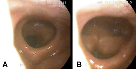
Figure 3
Images from follow-up flexible fiberoptic nasolaryngoscopy showing supraglottic stenosis with adherence of the lateral portions of the epiglottis to the posterior pharyngeal wall. (a) Normal arytenoid and vocal fold motion was noted with complete abduction (b) and adduction.
Discussion
Our case illustrates the potential for airway damage secondary to intranasal acetaminophen abuse. This young woman caused destruction of her intranasal and pharyngeal anatomy, with resultant nasopharyngeal and supraglottic stenosis, requiring temporary gastrostomy tube feedings because of her abusive habit. Prior reports of intranasal drug abuse describe early use of oral antibiotics to treat a presumptive pharyngitis [6,7]. This highlights a delay in diagnosis, often due to patients withholding information. Patients who inhale medications through one nostril exclusively typically have unilateral disease, helping rule out most infectious etiologies and guiding the clinician toward a correct diagnosis. One may visualize crushed pill debris in the nasal cavity, or analysis of intranasal debris can confirm the suspicion of drug abuse.
Intranasal abuse of acetaminophen-hydrocodone or acetaminophen-oxycodone combination drugs is well described [4,7]. Most patients present with necrosis of the oronasal mucosa, with pill debris visible during periods of active use. In cases of extensive intranasal drug abuse, perforation of the nasal septum or erosion of nasopharyngeal structures may occur. While reviewing intranasal abuse of opioid-acetaminophen compounds, Houlton et al. [7] found 66% of patients in their series had septal perforations, yet Alexander et al. [4] reported 51% of patients having a septal perforation. A recent case report by Hardison et al. [8] describes a patient with severe nasal, nasopharyngeal, and oropharyngeal erosion secondary to intranasal acetaminophen use, without involvement of the larynx. Interestingly, cocaine-associated septal perforation was much less common, with an incidence ranging between 4-11% [9].
Diffuse involvement of the nasal mucosa may result in fungal infection, as seen in our patient. A pathologic diagnosis of invasive fungal sinusitis (IFS) was made in this case, however her clinical disease was considered distinctly different from IFS. Other reports have also indicated similar pathologic findings without complete clinical correlation. In a retrospective chart review by Vosler et al. [6], pathology and cultures revealed non-invasive fungal disease in 85.7% of patients abusing intranasal acetaminophen-hydrocodone. These patients did not respond to antifungal treatment; however, their infections resolved with cessation of intranasal drug abuse. It is hypothesized that tissue necrosis facilitates superficial saprophytic fungal growth. Houlton et al. [7] found that Candida albicans is the most common fungus found in intranasal drug-induced fungal rhinopharyngitis, followed by Aspergillus fumigatus and the bacterium Staphylococcus aureus.
The etiology of mucosal and structural necrosis in patients abusing medications, including cocaine, narcotics, as well as non-prescription medications, is incompletely understood. While the vasoconstrictive properties of cocaine are well known and documented, similar patterns of necrosis may be seen in patients who abuse narcotics without these properties [4,6]. Multiple theories have been put forward, including osmotic properties of the medications and pills, adulterants in illicit drugs, as well as fillers used in the manufacture of the pills themselves. In the case of acetaminophen, a study by Hart et al. [10] examined the localization of acetaminophen in a mouse tissue model. They found toxic doses of acetaminophen can collect not only in the hepatocytes and renal cells, but olfactory epithelium. Talc, commonly a filler in drug tablets, is known to cause a granulomatous reaction in respiratory epithelium, both in inhaled and injected forms [6,11]. Prior reports of intranasal opioid/acetaminophen combination tablets have found evidence of talcosis in the mucosa of some of these patients.
Conclusion
Without patient description of abusive behaviors, intranasal drug abuse diagnosis may be delayed or missed entirely. Patients presenting with nasal and nasopharyngeal abnormalities such as necrosis of the oronasal mucosa or nasal septal perforation, particularly with unilateral findings, should raise suspicion of intranasal drug abuse. While the direct etiology of the damage is debated, there is likely a combination of factors that lead to mucosal injury. While there are few reported cases of intranasal abuse of over the counter medications such as acetaminophen, the easy accessibility of these drugs may lead to increased incidence of use with similar clinical findings to the abuse of prescription and illicit drugs.
References
- Hernandez SH, Nelson LS. Prescription drug abuse: insight into the epidemic. Clin Pharmacol Ther. 2010; 88: 307-317.
- Substance Abuse and Mental Health Services Administration. Results from the 2010 National Survey on Drug Use and Health: Summary of National Findings. 2011.
- National Institute on Drug Abuse (NIDA). Prescription Drugs & Cold Medicines. 2015.
- Alexander D, Alexander K, Valentino J. Intranasal hydrocodone-acetaminophen abuse induced necrosis of the nasal cavity and pharynx. Laryngoscope. 2012; 122: 2378-2381.
- IMS National Sales Perspectives, Year 2004-2008.
- Vosler PS, Ferguson BJ, Contreras JI, Wang EW, Schaitkin BM, Lee S. Clinical and pathologic characteristics of intranasal abuse of combined opioid-acetaminophen medications. Int Forum Allergy Rhinol. 2014; 4: 839-844.
- Houlton JJ, Donaldson AM, Zimmer L, Seiden A. Intranasal drug-induced fungal rhinopharyngitis. Int Forum Allergy Rhinol. 2012; 2: 130-134.
- Hardison SA, Marcum KK, Lintzenich CR. Severe necrosis of the palate and nasal septum resulting from intranasal abuse of acetaminophen. Ear Nose Throat J. 2015; 94: E40-42.
- Deutsch HL, Millard DR. A new cocaine abuse complex. Involvement of nose, septum, palate, and pharynx. Arch Otolaryngol Head Neck Surg. 1989; 115: 235-237.
- Hart SG, Cartun RW, Wyand DS, Khairallah EA, Cohen SD. Immunohistochemical localization of acetaminophen in target tissues of the CD-1 mouse: correspondence of covalent binding with toxicity. Fundam Appl Toxicol. 1995; 24: 260-274.
- Scheel AH, Krause D, Haars H, Schmitz I, Junker K. Talcum induced pneumoconiosis following inhalation of adulterated marijuana, a case report. Diagn Pathol. 2012; 7: 26.