Case Report
Eosinophilic Ulcer of Oral Mucosa: A Case Report
Shalabh Rastogi1*, Soumick Ranjan Sahoo1 and Somdatta B Datta2
1Department of Ent, Tata Motors Hospital, Telco Colony, India
2Department of Pathology, Tata Motors Hospital, India
*Corresponding author: Shalabh Rastogi, Department of ENT, Tata Motors Hospital, Jamshedpur, Jharkhand, Pin 831004, India
Published: 16 Aug, 2016
Cite this article as: Rastogi S, Sahoo SR, Datta SB.
Eosinophilic Ulcer of Oral Mucosa: A
Case Report. Ann Clin Case Rep. 2016;
1: 1066.
Abstract
Eosinophilic ulcer of Oral mucosa is a rare, self-limiting and benign lesion commonly involves tongue, lips, buccal mucosa, palate, gingival and floor of the mouth. Clinically it can be confused with malignant ulcer. We present the case of a 35-year-old male with a history of a painful ulcer on the right lateral side of his tongue treated surgically with excision biopsy. Histopathological examination showed feature suggestive of EUOM with no cellular atypia. The lesion showed full blown recurrence after excision, later healed itself with conservative management.
Introduction
Eosinophilic ulcer of the oral mucosa (EUOM) is considered to be a reactive lesion with benign clinical course occurs most commonly in 30 – 50 years of age. Although injury has been considered to play a major role, the exact etiology still remains obscure [1,2]. Predominantly lesions occur on the tongue but can occur at other sites like lips, buccal mucosa, palate, gingiva and floor of the mouth [2-4]. EUOM clinically manifested as rapidly developing solitary ulcer, with elevated and indurated borders [2] mimicking malignant ulcer. Microscopically, there is polymorphic inflammatory reaction rich in eosinophils extending deep into the submucosa, underlying muscle and salivary glands hence got its name of eosinophilic ulcer. EUOM is usually a self-limiting disorder that resolves spontaneously in few weeks [3]. Various treatment options reported in literature ranging from conservative to surgical excision of the lesion. Recurrence is extremely rare after excision [5]. We present a case of this uncommon disease which behaved differently from our expectations.
Case Presentation
A 35-year-old male patient presented to ENT outpatient department with chief complaint of
painful ulcer in the right lateral margin in the anterior 2/3 of tongue for past 2-month. Lesion was
progressively increasing and patient had difficulty in chewing. Patient was nonsmoker and did not
consume tobacco in any form. Patient had a jagged tooth which caused repeated trauma over tongue
resulting into ulcer. Patient had taken medication from other centers in the form of multivitamins,
steroidal ointments, oral steroids but lesion kept on increasing. There was no other significant past
medical history. On oral examination there was a small, firm, solitary ulcerative lesion measuring
1x1cm with uneven surface and induration. The base appeared yellowish white in color and freely
mobile over tongue muscles. The edges were raised and firm. The surrounding area showed redness.
There were no enlarged lymph nodes. A provisional diagnosis of non-healing ulcer of tongue was
considered. An excisional biopsy was done under local anesthesia removing complete ulcer taking
2 mm margin all along. Ulcer was excised completely till muscular layer of tongue. After excision
and securing hemostasis, wound was closed primarily with vicryl suture. Patient was asked for tooth
grinding for his jagged tooth. Histopathology showed features suggestive of eosinophilic ulcer of
tongue.
Patient showed full blown recurrence (Figure 1) within 3 weeks after excision. Since
histopathology showed no signs of malignancy and eosinophilic ulcers do respond to conservative
management, patient was again given a trial of conservative treatment before going for wide field
lesion excision which include steroidal ointment, multivitamins, antibiotic to clear any post-surgical
infection which may prevent healing, and this time the lesion responded and got completely healed
within next 14 days. Healthy tongue after complete resolution of ulcer shown in Figure 2.
Figure 1
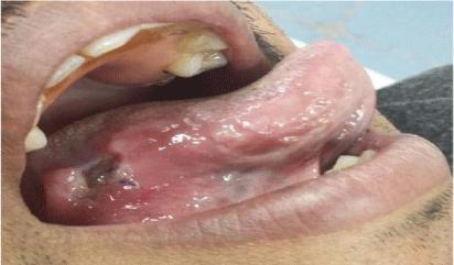
Figure 1
Recurrence of eosinophilic ulcer on right lateral margin of tongue after surgical excision showing yellow base with raised margins and remnant of vicryl suture in situ.
Figure 2
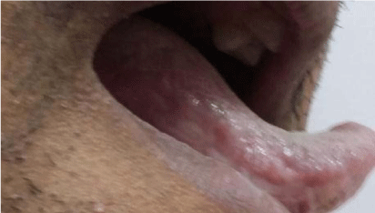
Figure 2
Completely healed ulcer after conservative management.
Figure 3

Figure 3
High power magnification showing inflammatory infiltrate rich in eosinophils extending into submucosa and muscle layer.
Discussion
Eosinophilic ulcers of tongue are relatively rare conditions and there is limited study available
about this clinical entity. Antonio Riga and F. Fede were among the first to describe this clinical entity in children hence it is also called Riga–Fede disease. The first description of this disease in adults was given by Popoff in 1956.
Though exact etio-pathogenesis is still controversial these lesions are usually of traumatic origin caused by repeated injury from sharp, fractured teeth or ill-fitting prosthetic material. Many others factors which had been implicated are infection, malnutrition, febrile states, hormonal stimulation, hereditary transmission of a dominant autosomal gene osteoblastic activity inside the tooth germ and hypovitaminosis [6]. Velez et al. [7] hypothesized that trauma cause’s easy access of virus and toxic agents to enter the underlying tissue and cause an inflammatory response led to the development of eosinophilic ulcer. In our case a jagged tooth was present which caused repeated trauma of tongue and led to ulcer formation but otherwise patient was healthy with no other systemic illness or malnutrition.
According to Velez pain is the main symptoms for which patient seek medical advice [7]. Our case was also presented as painful indurated ulcerative lesion of tongue with whitish yellow base not adherent to underlying muscles of tongue without any regional lymphadenopathy. These features are similar to features of EUOM as described in literature [1,3,8]. The differentials of EUOM to be considered are traumatic ulcers, squamous cell carcinoma, lymphoma, histiocytosis-x, tuberculosis, syphilis, herpes, histoplasmosis, discoid lupus erythematosus, and wegners granulomatosis [9]. In our case with evident history of tooth bite and no other systemic illness, differential diagnosis of non-healing traumatic ulcer and malignant ulcer was made. Lesion was subjected to excisional biopsy for histopathological confirmation.
Histopathological examination differentiates EUOM from malignancy. Characteristic features of eosinophilic ulcer include polymorphic inflammatory infiltrate extending deep into the submucosa, underlying muscle and salivary glands. In the background of mixed inflammatory reaction there is massive eosinophilia with large histiocytic cells with pale nuclei showing frequent mitoses, in some instances showing a pseudo-lymphomatous aspect [4,8]. Others components of the infiltrate include lymphocytes, plasma cells, granulocytes and mast cells and occasionally on microscopic examination it is misdiagnosed as lymphoma [10]. Figure 3 shows the same microscopic features present in our case with eosinophils counting as about 30% of total cell population.
Another such eosinophilic infiltration is seen in eosinophilic esophagitis whose pathogenesis is entirely different from EUOM. Microscopically also eosinophilic infiltration (> 15 eosinophils /hpf) of squamous mucosa apart from degranulated eosinophils, eosinophilic microabcesses and eosinophilic infiltrate in superficial layers of squamous mucosa constitute histologic findings in eosinophilic esophagitis. It never goes down till muscular layer in esophageal tissue [11].
Many authors have advocated wide range of treatment like wait and watch, antibiotic, topical steroid, Intralesional steroid, curettage, cryotherapy and surgical excision. No further local recurrences were usually noted after excision [5]. Segura and Pujol [3] reported new lesions formation at other mucosal sites. In our case lesion was excised for biopsy later to redevelop into similar looking ulcer in just 2-3 weeks’ time. With treatment of jagged tooth and malignancy thoroughly ruled out patient was again kept on conservative treatment before planning for wide field excision and patient responded by decrease in size of ulcer. The lesion completely disappears in next 3-week time.
This is an abnormal and unusual behavior shown by any benign lesion. Hence we conclude that if the histopathological report rules out malignancy we should wait and try for conservative management before going for wide field lesion excision.
References
- Ada S, Seckin D, Tarhan E, Buyuklu F, Cakmak O, Arikan U. Eosinophilic ulcer of the tongue. Australas J Dermatol. 2007; 48: 248-250.
- Ribeiro AL, Mendes FR, Alves Sde M Jr, Pinheiro Jde J. Eosinophilic ulcer: the role of stress-induced psychoneuroimmunologic factors. Oral Maxillofac Surg. 2011; 15: 179-182.
- Segura S, Pujol RM. Eosinophilic ulcer of the oral mucosa: a distinct entity or a non-specific reactive pattern? Oral Dis. 2008; 14: 287-295.
- Chatzistamou I, Doussis-Anagnostopoulou I, Georgiou G, Gkilas H, Prodromidis G, Andrikopoulou M, et al. Traumatic ulcerative granuloma with stromal eosinophilia: report of a case and literature review. J Oral Maxillofac Surg. 2012; 70: 349-353.
- Nagarajana NP, Somub L, Padmavathya PK, Sundarama S. Eosinophilic ulcer of the tongue - a rare and distinct entity. Sri Ramachandra Journal of Medicine. 2013; 6: 16-18.
- Ghimire N, Maharjan IK, Kumar A, Koirala B. Riga-Fede Disease. Health Renaissance. 2013; 11: 174-176.
- Vélez A, Alamillos FJ, Dean A, Rodas J, Acosta A. Eosinophilic ulcer of the oral mucosa: report of a recurrent case on the tongue. Clin Exp Dermatol. 1997; 22: 154-156.
- Nikitakis NG, Brooks JK. Persistent tongue ulcer. Eosinophilic ulceration. Gen Dent. 2010; 58: 150, 153.
- Raviteja YS, Kundana I, Triveni T. Eosinophilic Ulcer of the Oral Mucosa. Int J Dent Med Res. 2015; 1: 112-115.
- Shapiro L, Juhlin EA. Eosinophilic ulcer of the tongue report of two cases and review of the literature. Dermatologica. 1970; 140: 242-250.
- Dellon ES. Eosinophilic esophagitis: diagnostic tests and criteria. Curr Opin Gastroenterol. 2012; 28: 382-388.