Case Report
Painful Blindness: A Diagnosis not to be Missed
García-García ME*, Azorín DG, Almonacid JJ and Dolado AM
Department of Neurology, Hospital Clínico San Carlos, Spain
*Corresponding author: María Eugenia García-García, Neurology Department, Hospital Clínico San Carlos, C/ Profesor Martín Lagos s/n, 28039 Madrid, Spain
Published: 08 Aug, 2016
Cite this article as: García-García ME, Azorín DG,
Almonacid JJ, Dolado AM. Painful
Blindness: A Diagnosis not to be
Missed. Ann Clin Case Rep. 2016; 1:
1059.
Abstract
A 62-year-old female was admitted with a history of left sided headache, mild proptosis and left ocular redness occurring during the last weeks. A brain MRI showed findings suggesting a carotid fistula. The cerebral angiogram highlighted an indirect Barrow type D left carotid-cavernous fistula which was treated with embolization. The following day, the patient complained of left retro-orbital pain and loss of vision. The examination showed the following findings (in the left eye): mydriatic nonreactive pupil, vision loss, conjunctivalchemosis and an intraocular pressure of 33 mmHg. The new angiogram showed an almost complete closure of the fistula and the brain MRI demonstrated a choroidal detachment along the medial wall of the left ocular globe which provoked acute angle closure glaucoma. Considering that an early diagnosis and treatment are crucial to the prognosis, neuro-ophthalmologists should consider this entity in the differential diagnosis of a patient with worsening ocular symptoms after a carotid-cavernous fistula embolization.
Keywords: Carotid-Cavernous fistula; Choroidal detachment; Angle closure glaucoma; Embolization; Angiography cerebral
Introduction
Carotid cavernous fistulas (CCFs) are anomalous connections between the carotid arterial
system and the cavernous sinus and they are normally classified in two main categories, depending
on the arterial system involved. The first group, direct or high flow shunts (type A), are characterized
by a direct connection between the internal carotid artery and one or more of the venous channels
within the sinus; the second group are indirect or low flow shunts, which arise from the dural
branches of internal, external carotid system or both (type B, C, D respectively) [1]. The symptoms
and signs produced by these lesions depend on several factors but the majority of them are ocular,
usually ipsilateral to the fistula. Endovascular embolization currently represents the first therapeutic
modality for CCFs because it is associated with high occlusion and low complication rates [2].
In this report, we describe a patient with loss of vision and acute angle closure glaucoma (AACG)
secondary to choroidal detachment, a very infrequent complication after carotid cavernous fistula
embolization.
Case Report
A 62-year-old female was admitted to emergency care with a history of left sided headache, mild
proptosis and left ocular redness occurring during the preceding four weeks. On admission, the
ophthalmologic examination revealed a subtle exophthalmos with epibulbar congestion and some
tortuous corkscrew-type blood vessels in the left eye (Figure 1). The visual acuity was 20/25 Oculus
Dexter (OD) and 20/20 Oculus Sinister (OS), with normal colour vision in the Ishihara pseudoisochromatic
plates. The pupils were isocoric and reactive to light and there were no limitations
in the ocular motility, ptosis nor bruits. The funduscopic examination showed slightly dilated
retinal veins and optic disc swelling without retinal hemorrhages in OS. Intraocular pressure was 18
mmHg OD and 23 mmHg OS. Medical history was significant only for hypertension and she denied
suffering any head trauma.
Laboratory tests, which included thyroid profiles such as T3, T4, TSH and TSH receptor antibody
were all within normal ranges. A cranial MRI and a 3D time-of-flight (TOF) MR angiography
revealed proptosis, an enlarged left superior ophthalmic vein (SOV) and a dilated left cavernous sinus
with multiple abnormal flow voids which filled in early during arterial phase. Digital subtraction
angiography (DSA) confirmed an indirect Barrow type D left carotid cavernous fistula originating
from both arteries, the right external carotid (through internal maxillary artery) and from the right
internal carotid (through meningo-hypophyseal trunk) (Figure 2A and B). No aneurysms were
identified. The patient underwent transarterial embolization with detachable coils resulting in an almost complete closure of the fistula and with no complications. Twenty four hours later she complained of moderate to severe retro orbital pain, loss of vision in the left eye, nausea and vomiting. At that moment, the ocular examination revealed amydriatic nonreactive left pupil, vision reduced to only light perception, limitation of horizontal ocular motility, conjunctivalchemosis, severe proptosis and an intraocular pressure of 33 mmHg (Figure 3). Gonioscopic findings demonstrated a closed angle in the OS with a normal OD anterior chamber angle. A new cerebral DSA was immediately performed showing little and early filling of the left sinus cavernous through a small meningo-hypophyseal trunk with no flow in the SOV, suggesting a possible thrombosis at that point (Figure 4). Given that these findings did not explain all the symptoms (such as severe loss of vision), a cranial MRI was carried out which revealed a left thrombosed SOV as well as a choroidal detachment along the entire medial wall of the left ocular globe, extending up to the ciliary body and provoking its anterior displacement and consequently an acute angle closure glaucoma (Figure 5). Topical instillation of 0.5% timolol maleatedrops, 750 mg of oral acetazolamide per day and left canthotomy-cantholysis were necessary to control intraocular pressure, which lowered to 17 mmHg the following day. At a three month follow-up visit, the patient had improved significantly with no ocular symptoms and her visual acuity was 20/25 OS. A new DSA showed a complete closure of the CCF.
Figure 1
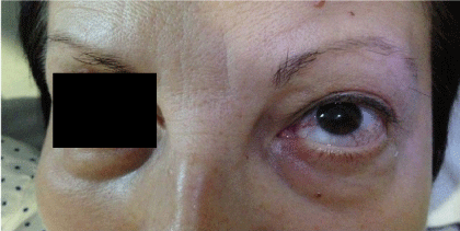
Figure 1
External photograph showing a mild exophthalmos, redness and dilated episcleral vessels in the left eye.
Figure 2a

Figure 2a
Right internal carotid angiogram showing an indirect Barrow type D left carotid cavernous fistula. Early filling of the left cavernous sinus (arrow) fed by branches from the right internal carotid (through meningo-hypophyseal trunk).
Figure 2b
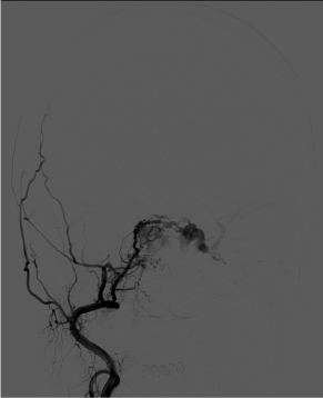
Figure 2b
The dorsal arch height change measured following the application of the tape.
Figure 3
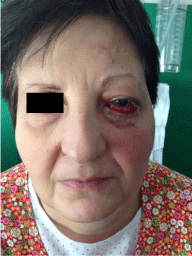
Figure 3
External photograph (front view) showing a clinical worsening with proptosis, ocular redness, conjunctivalchemosis occurring 24 hours after embolization of left CCF.
Discussion
Endovascular procedures involving the use of different embolic materials are one of the main therapeutic interventions for CCFs, with success rates between 70-78% for closing indirect carotid cavernous fistulas [2]. Different complications can be present up to 5% of the procedures, being iatrogenic choroidal detachment and subsequently acute angle closure glaucoma (AACG) an extremely rare complication and no more than a few cases have been described in the literature. The majority of them developed several hours after the embolization (as in our patient) however, AACG can also occur after angiography and endovascular manipulation without embolizationand may even develop several weeks after the intervention [3-5]. The pathophysiological hypothesis is unclear but three different mechanisms have been proposed: 1) the hemodynamic changes produced by the CFF itself (venous stasis and orbital congestion); 2) the SOV thrombosis due to the embolization and 3) the absence of sufficient collateral drainage. All together could lead to increased pressure and permeability in the vortex veins, transudation into the supra choroidal space (normally virtual) and a choroidal separation. Subsequently, the detachment produced a forward shift of the iris-lens diaphragm, closing of the anterior chamber with failure of the aqueous humor circulation and finally, an AACG [4-6]. The worsening in our patient occurred over night, in the darkness, which favors pupil dilation, thus further narrowing the angle and increasing the propensity to develop an AACG. Neuro imaging techniques, such as brain MRI, can help us in the differential diagnosis showing the choroidal detachment as a semilunar or ring shaped area of increased signal on both T1and T2 weighted images [7].
Considering that an early diagnosis and treatment are crucial to avoid deterioration of choroidal and retinal circulation, neuroophthalmologists should consider this entity in the differential diagnosis of a patient with vision loss after CCF embolization. Medical management, canthotomy-cantholysis, iridotomy, choroidal drainage are useful treatments to decrease intraocular pressure if the CFF has been previously closed.
Figure 4
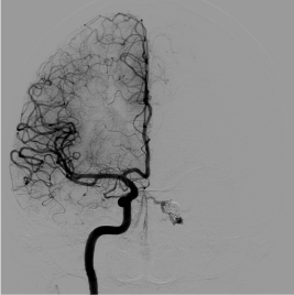
Figure 4
Right internal carotid angiogram (front view) shows little and early filling of the left cavernous sinus through a small meningo-hypophyseal trunk.
Figure 5
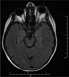
Figure 5
Axial Flair MRI image showing the choroidal detachment along medial wall of the left ocular globe extending up to the ciliary body (arrow).
References
- Barrow DL, Spector RH, Braun IF Landman JA, Tindall SC, Tindall GT. Classification and treatment of spontaneous carotid-cavernous sinus fistulas. J Neurosurg. 1985; 62: 248-256.
- Gemmete JJ, Ansari SA, Gandhi DM. Endovascular techniques for treatment of carotid cavernous fistula. J Neuroophthalmol. 2009; 29: 62-71.
- Golnik KC, Newman SA, Ferguson R. Angle-closure glaucoma consequent to embolization of dural cavernous sinus fistula. AJNR Am J Neuroradiol. 1991; 12: 1074-1076.
- Talajic JC, Assalian A, Roy D, Harasymowycz PJ. Angle closure glaucoma after angiography of carotid cavernous fistula. J Glaucoma. 2010; 19: 73-74.
- Thinda S, Melson MR, Kuchtey RW. Worsening angle closure glaucoma and choroidal detachments subsequent to closure of a carotid cavernous fistula. BMC Ophthalmology. 2012; 12: 28.
- Komiyama M, Morikawa T, Matsusaka Y, Yasui T, Shimizu H. Acute angle-closure glaucoma after successful embolization of traumatic carotid cavernous fistula. Neurol Med Chir (Tokyo). 2003; 43: 142-145.
- Mafee MF, Peyman GA, Tan WS. Magnetic resonance imaging and computed tomography of choroidal detachment. Acta Radiol Suppl. 1986; 369: 327-330.