Case Report
Amebic Liver Abscess Masquerading Like Pyogenic Hepatic Abscess: Get the Anchovy Sauce
Lee BT* and Kaplowitz N
Department of Gastrointestinal and Liver Diseases, Keck School of Medicine of USC, USA
*Corresponding author: Brian T Lee, Division of Gastrointestinal and Liver Diseases, Department of Medicine Keck School of Medicine of USC, 1200 N State St. Los Angeles, CA 90033, USA
Published: 21 Jul, 2016
Cite this article as: Lee BT, Kaplowitz N. Amebic Liver
Abscess Masquerading Like Pyogenic
Hepatic Abscess: Get the Anchovy
Sauce. Ann Clin Case Rep. 2016; 1:
1050.
Abstract
Amebic liver abscess is an extra intestinal manifestation of an infection from the protozoan, Entamoeba histolytica. On the contrary, pyogenic hepatic abscess is a result of bacterial infection stemming from the bloodstream, biliary tract, abdominal cavity, or portal system. We report a case of a patient with an amebic liver abscess initially presenting as a pyogenic hepatic abscess. A 66-yearold man presented with three days of malaise, cough, and right flank pain. Initial Chest CT revealed two large lesions within the right hepatic lobe. A percutaneous drain of the inferior lobe abscess emptied thick anchovy paste-like material. E. histolytica IgG serology returned positive. He was maintained on metronidazole with the addition of diiodohydroxyquin for adjuvant luminal therapy. Although treatment is distinctive for each individual disease, it is prudent to initiate treatment for both, especially in the early management of severe illness until the diagnosis is confirmed.
Keywords: Amebic liver abscess; E. histolytica; Protozoan; Pyogenic hepatic abscess; Hepatomegaly
Abbrevations: ALA: Amebic Liver Abscess; PHA: Pyogenic Hepatic Abscess; AST: Aspartate Amino Transferase
Introduction
Amebic liver abscess (ALA) is an extra intestinal manifestation of an infection from the protozoan, Entamoeba histolytica. Most commonly, amebiasis can seed through the portal tract and result in the formation of a liver abscess [1]. Initial presentation is significant for abdominal pain and hepatomegaly. Diagnosis is made with negative blood cultures but positive amebic serology. On the contrary, pyogenic hepatic abscess (PHA) is a result of bacterial infection stemming from the bloodstream, biliary tract, abdominal cavity, or portal system. It commonly presents as fever, chills, nausea, vomiting, and often, jaundice. Diagnosis is made with positive blood and/or abscess cultures. We report a unique case of a patient with a massive ALA initially presenting as PHA.
Case Presentation
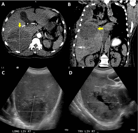
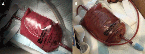
A 66-year-old Hispanic man was well until he presented with three days of malaise, productive cough, and right flank pain. Initial examination was notable for fever (temperature 101.3 degrees Fahrenheit), tachycardia, tachypnea, tender hepatomegaly, encephalopathy and jaundice. Blood work was remarkable (sodium 114 mmol/L, bicarbonate 17 mmol/L, alkaline phosphatase 488 U/L, albumin 2.4 g/dL, aspartate amino transferase (AST) 241 U/L, alanine transaminase 208 U/L, total bilirubin 5.1 mg/dL, direct bilirubin 4.0 mg/dL, lactate 5.5 mmol/L, white blood cell count 57.7 x 10³ with 97% neutrophils, hemoglobin 11.1 g/dL, platelets 65 x 103, INR 1.62). Upon presentation, he was intubated, admitted to the intensive care unit, and initiated on intravenous antibiotics with vancomycin, cefepime, and metronidazole. Initial Chest CT revealed two large non-arterially enhancing, ill-defined, hypo attenuating lesions within the right hepatic lobe, the largest measuring up to 11.7 cm (Figure 1). A followup abdominal ultrasound revealed two hypoechoic lesions with hyperechoic rims; the first lesion measured 10 x 9.3 x 9.2 cm while the second measured 11.2 x 8.2 x 10.7 cm (Figure 1). Infectious Disease was consulted and antibiotics were switched to vancomycin, meropenem and metronidazole to cover for other etiologies of hepatic abscesses. Initial blood cultures were negative. Stool analysis was negative for infection. A percutaneous drain of the inferior lobe abscess emptied 600 milliliters of thick anchovy paste-like material (Figure 2). Gram stain and culture of this aspirate were negative. He had another drain placed into the superior lobe abscess, which appeared to rupture into the pleural space; both drains were subsequently extracted after adequate drainage. He was extubated after clinical improvement on antibiotics. Eventually, E. histolytica IgG serology returned positive, and metronidazole alone was continued. The right-sided pleural effusion secondary to a rupture of the abscess was managed successfully with serial drain placements. He was maintained on metronidazole for 10 days with the addition of diiodohydroxyquin for adjuvant luminal therapy for a total of 21 days.
Figure 1
Figure 1
Body imaging with chest CT and abdominal ultrasound. (A) CT image, axial view. Lesion seen in right lobe. (B) CT image, coronal view. Note the elevation of the right hemi diaphragm and pleural effusion. (C) US image, longitudinal view. (D) US image, transverse view. Note the central cystic appearance.
Figure 2
Discussion
While ALA is predominant in males, it is usually a disease of younger adults when compared to PHA [2]. Several case series have reported that those in their fifth decade or older were more consistent with PHA [3]. Our patient was a male in his sixth decade of life. PHA is associated with variable complaints, such as cough or chest pain [2]. Our patient did have complaints of a productive cough. Lab abnormalities are more noteworthy in PHA, particularly hyperbilirubinemia, which is rare in ALA [3]. Other lab abnormalities more typical of PHA include elevated lactic dehydrogenase, low albumin, increased AST, and leukocytosis with a marked shift to the left (>89% neutrophils) [2]. However, as illustrated in our case, the distinction between ALA and PHA can be very challenging and aspiration of the abscess is needed both for diagnosis and for prevention of rupture of strategically located abscess. Definitive diagnosis of ALA is by demonstrating amebic trophozoites in the aspirate of the liver abscess. Due to the cellular response to invading trophozoites occurring mostly in the periphery of the lesion, a microscopic evaluation may be low yield due to imprecise sampling. Aspirate evaluation has higher yield in PHA where microorganisms and polymorphonuclear cells are more evident. The fluid aspirate in this case was not tested for trophozoites but rather for gram stain and cultures in order to definitively rule out PHA. Given the low yield of fluid aspirate analysis, serologies for E. histolytica remain instrumental in the diagnosis and differentiation of ALA from PHA. The ELISA remains the most sensitive assay in detecting circulating antibodies specific for E. histolytica. It is important to note that serology may not be apparent in the first 7-10 days of infection [1]. Although treatment is distinctive for each individual disease, it is prudent to initiate treatment for both, especially in the early management of severe illness until the diagnosis is confirmed.
References
- Salles JM, Salles MJ, Moraes LA, Silva MC. Invasive amebiasis: an update on diagnosis and management. Expert Rev Anti Infect Ther. 2007; 5: 893-901.
- Barnes PF, De cock KM, Reynolds TN, Ralls PW. A comparison of amebic and pyogenic abscess of the liver. Medicine (Baltimore). 1987; 66: 472-483.
- Cosme A, Ojeda E, Zamarreño I, Bujanda L, Garmendia G, Echeverría MJ, et al. Pyogenic versus amoebic liver abscesses. A comparative clinical study in a series of 58 patients. Rev EspEnferm Dig. 2010; 102: 90-99.


