Case Report
Unusual Presentation of Hodgkin Lymphoma with Limbic Encephalitis (Ophelia Syndrome)
Ramachandran P* and O’Brien T
Department of Hematology/Oncology, Metrohealth Medical Center, USA
*Corresponding author: Ramachandran P, Department of Oncology, Metrohealth Medical Center, Cleveland, Ohio 44104, USA
Published: 15 Jul, 2016
Cite this article as: Ramachandran P, O’Brien T. Unusual
Presentation of Hodgkin Lymphoma
with Limbic Encephalitis (Ophelia
Syndrome). Ann Clin Case Rep. 2016;
1: 1047.
Abstract
Introduction: Hodgkin lymphoma is a potentially curable lymphoma which has unique biological,
clinical and histological characteristics. It is associated with multiple paraneoplastic neurological
syndromes of which limbic encephalitis is a very rare presentation. Antibody against mGLUR5
seems to be associated for this presentation.
Case Presentation: Here we describe a rare case of a 60 year old Caucasian lady newly diagnosed
with stage 4B Hodgkin lymphoma who presented with progressive neurological deterioration.
Timely decision to treat her underlying malignancy with chemotherapy resulted in are markable
recovery in her neurological status within few weeks of treatment.
Conclusion: This case emphasis the need to investigate for paraneoplastic disorders when patients
with Hodgkin lymphoma presents with neurological changes. Sometimes it may be the only
presentation of the underlying tumor which poses a diagnostic challenge to the physicians. Early
treatment of the underlying tumor helps in successful neurological recovery.
Keywords: Ophelia syndrome; Limbic encephalitis; Hodgkin lymphoma
Introduction
Paraneoplastic disorders in Hodgkin lymphoma is a very rare presentation. It poses a diagnostic challenge when this may be the only presentation of the underlying tumor. Here we discribe a rare case of 60 year old caucasian lady with newly diagnosed stage 4B Hodgkin lymphoma who presented with worsening neurological deterioration of unclear etiology. Paraneoplastic syndrome due to Hodgkin lymphoma was suspected and the patient was treated with chemotherapy. Within a few weeks of treatment, the patient improved neurologically and was back to her baseline. She completed her chemotherapy and her disease is currently in remission.
Case Presentation
The patient is a 60 year old Caucasian female who presented to a tertiary facility with symptoms
of cough, SOB, weight loss and pruritic lesions over her body. On presentation she was febrile,
had diffuse excoriations all over her body and a palpable lymphadenopathy in her groin. She had
evidence of poor air entry in her chest and non-tender abdomen with hyperactive bowel sounds.
Neurologically she was alert but seemed to be slightly confused. Her laboratory abnormalities
included normocytic anemia with a Hb of 7.4, leukocytosis with WBC count of 25, thrombocytopenia
with platelet count of 99, INR of 1.4 and acute renal impairment with creatinine of 1.76. Her
chest x-ray showed left basilar atelectasis and her CT chest/abdomen/pelvis showed mediastinal,
abdominopelvic, retroperitoneal lymphadenopathy and splenomegaly. The largest lymph node was
para-aortic lymph node measuring 3.2 cm (Figures 1-4).
A careful medical history revealed hypertension, hyperlipidemia and oxygen dependent COPD
(FEV1-36%). Her drug history included statin, lisinopril, albuterol and ipratropium inhalers. Her
family history was significant for liver cancer in her father, lung cancer in her uncle and liver cancer
in her aunt. She is also an ex-smoker with a 45 pack year history and she quit smoking 8 years ago.
The patient was initially treated with broad spectrum antibiotics for a community acquired
pneumonia. She was also supported with packed red cell transfusion for her anemia and IV fluids
for ARF. She required admission to intensive care unit due to labile blood pressures and high
oxygen requirement. She required cardio respiratory support with ventilator and vasopressors. She
was further investigated with an ultrasound guided right inguinal lymph node biopsy which was consistent with Hodgkin lymphoma. A staging bone marrow biopsy also showed evidence of bone marrow involvement of Hodgkin lymphoma. Hence she had Classic Hodgkin Lymphoma stage 4B. Her skin lesions were also biopsied but were consistent only with excoriations (Figure 5 and 6).
Her clinical course was further complicated by deterioration in her mental status as she became comatose within a week into admission. She was investigated extensively with LP, CT head, MRI head and an EEG but none revealed a cause. During this work up she had evidence of mild vitamin B12 deficiency which was replaced.
Since the cause of her neurological deterioration was unclear, we considered the possibility of limbic encephalitis, a paraneoplastic syndrome in Hodgkin lymphoma. According to literature [1], limbic encephalitis in patients with Hodgkin Lymphoma has a better prognosis, as successful treatment of the tumor is sufficient to result in full neurological recovery. Hence we decided to treat her with chemotherapy. Anti-mGluR5 antibodies unfortunately could not be tested. She was treated with AVD (Adriamycin, vinblastine and Dacarbazine). Bleomycin was omitted due to her increased oxygen requirements. She had to be supported with CVVH during her initial chemotherapy cycle due to the risk of tumourlysis syndrome in the setting of anuric renal failure.
She started showing some neurological improvement after a week of chemotherapy. Her multi-organ damage gradually started to improve and within 2 weeks she was independent of ventilatory and vasopressor supports. She made a complete neurological recovery within 6 weeks and her mental status returned to baseline. She was finally discharged home after completing 2 cycles of chemotherapy.
She later completed further 4 cycles of chemotherapy on an outpatient basis and successfully remains in remission currently.
Figure 1
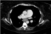
Figure 1
CT chest/abdomen/Pelvis showing mediastinal region.
Figure 2
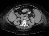
Figure 2
CT chest/abdomen/Pelvis showing abdominopelvic region.
Figure 3
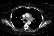
Figure 3
CT chest/abdomen/Pelvis showing retroperitoneal lymphadenopathy.
Figure 4
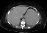
Figure 4
CT chest/abdomen/Pelvis showing splenomegaly.
Figure 5
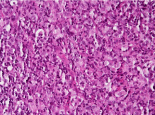
Figure 5
LN section revealing mixture of small lymphocytes, few neutrophils, plasma cells and scattered large atypical cells with prominent nucleoli, some binucleated suggestive of Reed Sternberg cells.
Figure 6
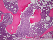
Figure 6
BM biopsy showing patchy granuloma like infiltrate with many large atypical cells with prominent nucleoli consistent with Reed Sternberg cells.
Discussion
Hodgkin lymphoma is a potentially curable lymphoma, primarily of B cell lineage that has unique histological, biological, immunophenotypical and clinical characteristics. Neurological complications of Hodgkin lymphoma can be separated into those that result directly from the disease or indirectly from the disease or from its treatment. Paraneoplastic disorders in Hodgkin lymphoma include cerebellar degeneration or limbic encephalitis which is rare presentations.
LE is also caused by is also caused by SCLC, breast cancer and testicular germ cell tumors [2]. Hodgkin Lymphoma is the third common cause of LE. The clinical and MRI features of LE in all these cases are same although the LE of HL patients has a better prognosis and successful treatment of the tumor is sufficient to result in full neurological recovery. This is probably related to the finding that this type of LE occurs in association with an antibody against the mGluR5 [3] that probably results in reversible neuronal dysfunction rather than neuronal death [1]. Unlike HL, LE is extremely rare in patients with NHL, with only a few cases described mostly in Japanese literature [4].
In 1982, Ian Carr4described an unusual neuropsychiatric disorder in his teenage daughter who had Hodgkin lymphoma presenting with progressive loss of memory, depression, hallucination and bizarre behavior. Dr. Carr postulated that a circulating neurotransmitter-like molecule mGluR5 antibody secreted by the tumor caused the disorder, which he named ‘Ophelia Syndrome’.
Our patient's case fulfilled all the criteria required for diagnosis of Ophelia syndrome [5] which included a compatible clinical presentation including neuropsychiatric symptoms due to involvement of limbic system; an interval of less than 4 years between the onset of neurological symptoms and diagnosis of HL; exclusion of other causes for limbic encephalitis; improvement of neurological symptoms with chemotherapy.
Given that HL has a bimodal age distribution and Ophelia syndrome usually precedes the diagnosis of HL, mGluR5 antibody needs to be considered in all patients presenting with limbic encephalitis of unclear etiology. The main concern of physician in those cases is to identify this condition and investigate for HL.
There has been at least 13 cases [6] of Ophelia syndrome reported so far. Of those 13 patients, 7 patients were tested for mGluR5 antibodies and 2 patients had positive results. Among these 13 patients, 12 received treatments with either chemotherapy or radiotherapy. Eight had full neurological recovery, 3 did not improve, 1 had partial improvement and the response of one patient is unknown.
Conclusion
This patient’s case demonstrates an extremely rare presentation of Hodgkin lymphoma as well as the better prognosis of the paraneoplastic presentation if treatment of the underlying tumor is instituted early. Paraneoplastic limbic encephalitis (Ophelia syndrome) should be considered in all patients with Hodgkin lymphoma who presents with mental state changes, since chemotherapy may dramatically affect the outcome.
References
- Mat A, Adler H, Merwick A, Chadwick G, Gullo G, Dalmau JO, et al. Ophelia syndrome with metabotropic glutamate receptor 5 antibodies in CSF. Neurology. 2013; 80: 1349-1350.
- Honnorat J, Antoine JC. Paraneoplastic neurological syndromes. Orphanet J Rare Dis. 2007; 2: 22.
- Carr I. The Ophelia syndrome: memory loss in Hodgkin's disease. Lancet. 1982; 1: 844-845.
- Dögel D, Beuing O, Koenigsmann M, Diete S. [Paraneoplastic limbic encephalitis resulting from non-Hodgkin-lymphoma: two case reports]. Fortschr Neurol Psychiatr. 2008; 76: 41-46.
- Olmos D, Rueda A, Jurado JM, Alba E. Presentation of Hodgkin Lymphoma with Ophelia Syndrome. American Journal of Clinical Oncology. 2007: 25: 1802-1806.
- Graus F, Ariño H, Dalmau J. Paraneoplastic neurological syndromes in Hodgkin and non-Hodgkin lymphomas. Blood. 2014; 123: 3230-3238.