Case Report
Primary Bone Lymphoma in a Patient Presenting with Persistent Knee Pain: A Case Report
Wills BW1*, Momaya AM1 and Wicker J2
1Department of Orthopedics, University of Alabama at Birmingham, USA
2Department of Pathology, University of Alabama at Birmingham, USA
*Corresponding author: Wills BW, University of Alabama at Birmingham, 1313 13th Street South Birmingham Alabama 35213, USA
Published: 15 Jul, 2016
Cite this article as: Wills BW, Momaya AM, Wicker J.
Primary Bone Lymphoma in a Patient
Presenting with Persistent Knee Pain: A
Case Report. Ann Clin Case Rep. 2016;
1: 1043.
Abstract
Primary lymphoma of bone is an uncommon bone disease typically occurring in the pelvis, spine, and ribs. Only five cases have been reported as beginning in the patella. Here, we report a case of a patient who presented with persistent knee pain one year after a patellar fracture. After an inconclusive CT-guided biopsy, he underwent an open patellar biopsy with frozen section and curettage and grafting. The pathology was consistent with primary lymphoma of the patella. We review the patient’s workup and treatment course.
Keywords: Bone lymphoma; Knee pain
History and Treatment Course
Prior to case write-up, written informed consent was obtained from our patient for the print
and electronic publication of his case, including text regarding history, physical, diagnostic findings
and workup, and all radiography/histology. Consent was also given for any reprinting in foreign
editions.
A 71-year-old Caucasian male presented to the orthopedic oncology clinic with persistent
right knee pain one year after sustaining a fracture of the patella. The fracture was initially treated
conservatively with an air cast and bracing by an outside hospital. The patient complained of
continued pain and swelling in his knee, but had no calf pain. Range of motion of the knee was
limited to 25-120 degrees. There were no palpable soft tissue masses, however plain radiographs
showed erosive changes in the patella and a possible adjacent soft tissue mass. Further workup was
ordered, including a right knee magnetic resonance imaging (MRI), with contrast, and a CT-guided
biopsy of the right patella (Figure 1).
The CT-guided biopsy was read as inconclusive by pathology. The MRI was reviewed and
confirmed erosive changes in the patella with abnormal signal changes (Figure 2). At this time,
we decided to proceed with further diagnostic workup. The patient underwent open biopsy of the
patella with frozen section, curettage and iliac crest grafting. The patient remained in the hospital for
standard postoperative antibiotics and was discharged postoperative day one.
Pathologic examination of the patellar mass demonstrated direct invasion from the bone into
the soft tissue with a lymphocytic infiltrate composed of large atypical cells with irregular nuclei and
occasional apoptotic bodies (Figure 3). Immunohistochemical staining showed that the neoplastic
cells were diffusely and strongly positive for CD20 and CD79a expression, with a subset of cells
exhibiting positive staining for Bcl-6 and CD10. Immunohistochemical staining for Ki-67 showed a
proliferative index of approximately 60%. These pathologic findings were consistent with a diagnosis
of diffuse large B cell lymphoma (DLBCL), germinal center subtype. A follow-up positron emission
tomography (PET) scan showed no other sites of involvement.
The patient was referred to medical oncology for further treatment. He subsequently underwent
successful chemotherapy without local radiation. At his one year follow-up clinic visit, the patient
was doing well and had completed all therapy. His chronic pain had resolved and no recurrence
or metastases were found. Additionally, the range of motion of his right knee returned to that
symmetric to the contralateral knee.
Figure 1

Figure 1
(A) AP, (B) lateral, and (C) sunrise views of the knee. The patella (white arrows) exhibits erosive osteolytic changes in all views, denoted by increased lucency compared to the surrounding bones. In Image A, the patella is outlined in white, and erosive (radiolucent) changes are notable at the superior pole (white arrow).
Figure 2

Figure 2
(A) Sagittal and (B) transverse T2-weighted magnetic resonance imaging (MRI) of the knee demonstrating heterogenous contrast enhancement of the patella (white arrows) and indicating erosive damage.
Figure 3
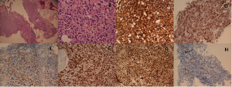
Figure 3
Histologic examination of the right patellar mass reveals fragments of bone and soft tissue with focal involvement of soft tissue by a lymphocytic infiltrate (A) composed of large atypical cells with irregular nuclei and occasional apoptotic bodies (B). Immunohistochemical staining shows that the neoplastic cells are strongly and diffusely positive for CD20 (C) and CD79a (D) expression. The cells of interest show focal weak staining for CD10 expression (E). The proliferative index is approximately 60% as indicated by Ki-67 expression (F). A predominant subset shows strong positive staining for Bcl-6 expression (G), Findings are consistent with a diagnosis of diffuse large B cell lymphoma (DLBCL), germinal center subtype while staining for MUM-1 highlights only rare positive cells (H).
Discussion
To our knowledge, primary lymphoma arising in the patella has only been previously reported in five cases [1-3]. Chandra and Eilender discussed a patient presenting with knee pain following a pop while walking that was discovered to be primary patellar lymphoma [3]. The patient was still in remission thirteen months following treatment. Additionally, two cases of patellar involvement in the setting of systemic lymphomatous disease have been reported [4,5]. Previous reports are limited by timeliness of diagnostic methods and incomplete case details such as histologic diagnosis or therapeutic effectiveness. Here, we seek to augment the literature by adding a comprehensive overview of a recent case of primary lymphoma of bone isolated to the patella.
Primary lymphoma of bone (PLB) accounts for 3-7% of all primary bone tumors and occurs in a wide spectrum of patients [6]. PLB commonly presents in the pelvis, spine and ribs. It also tends to occur in patients over 60 and in males slightly more than females [7,8]. The typical clinical presentation includes pain unrelieved by rest and warm, swollen soft tissue masses. Patients may report “B” symptoms such as fever of unknown origin or weight loss. Many patients will present with a pathologic fracture of the involved site and very rarely has it been described of primary bone lymphoma occurring following trauma [9,10]. Radio graphically, primary bone lymphomas are classically described aslytic, moth-eaten lesions with cortical thickening [11]. The most common PLB is Non-Hodgkin’s diffuse large B-cell lymphoma (DLBCL), as seen in our patient [8]. Approximately 18% of all lymphomas were non-specified DLBCL, 13% anaplastic large cell, 11.5% B-lymphoblastic lymphoma, 4.9% mantle cell lymphoma, 3.3% marginal zone lymphoma, 1.6% T-lymphoblastic lymphoma, 6.6 % Hodgkin lymphoma and 3.3% T cell origin lymphomas [11,12]. DLBCL accounts for 50-80% of primary bone lymphomas. Evidence of germinal center derivation, as in our case, is noted in approximately 50% of cases [12]. Histologically, DLBCL presents as mixed small round blue cells. Tumor cells are immunoreactive for CD45, CD20, and CD79awith variable immunoreactivity for CD10 and CD75 [12]. BCL-2 and BCL-6 immunoreactivity has been reported in 35% and 69% of cases, respectively [7].
Lesions presenting in the patella are rare and usually non-neoplastic [1]. When radiographic abnormalities of the patella are present, non-neoplastic conditions to consider include parathyroid disease, osteomyelitis, and gout. The most common tumors involving the patella are benign and include chondroblastomas and giant cell tumors [13]. The age of presentation assists in narrowing the differential diagnosis: giant cell tumor, chondroblastoma, aneurysmal bone cyst, osteomyelitis, osteoid osteoma, and solitary bone cyst occur more often in patients under 40. Intra-osseous gout, metastasis and intra-osseous ganglion occur more often in patients over 40 [13]. Radio graphically, soft tissue extension should raise the possibility of gout or a malignant lesion. However, it is important to note that PLB does not always have associated soft tissue involvement, and radio graphically may easily mimic a benign lesion of the patella [1].
Once a lymphomatous malignancy is confirmed histologically, it is imperative to ascertain the primary versus systemic nature of the disease and the extent of dissemination, as prognosis varies with tumor burden as well as histologic subtype. This staging process is most easily accomplished via full-body PET and/or CT scanning. Imaging should always be accompanied by a thorough history inquiring about systemic symptoms (e.g., classic “B” symptoms) and any other contributory aspects of the patient’s previous medical history (e.g., abnormal lab findings, genetic predisposition). Physical exam should address the presence or absence of palpable soft tissue masses and lymphadenopathy in all major anatomic regions.
Treatment for PLB typically involves chemotherapy, specifically with the CHOP regimen (cyclophosphamide, doxorubicin, vincristine, and prednisone) [14]. Adjunctive treatment with localized radiation is controversial [15], and orthopedic surgical intervention is usually only indicated for initial biopsy or pathologic fracture fixation. Long-term surveillance should also be considered in order to monitor for distant metastases or local relapse.
The rare nature of patellar PLB warrants increased awareness in order to minimize diagnostic delays. In our patient, it is difficult to discern whether the initial fracture of the patella was pathologic and therefore we cannot determine how long his condition went undiagnosed. It is possible that his initial trauma activated or triggered quiescent disease. Regardless, prognostic studies are currently scant; therefore, thorough follow-up should be encouraged of any physician who encounters a similar patient in the future.
Conclusion
Primary lymphoma of the patella is extremely rare. When patients present with persistent knee pain in the setting of a lesion, a pathologic diagnosis is essential. Patients should then be referred to an oncologist, if needed, for further treatment.
References
- Kransdorf MJ, Moser RP, Vinh TN, Aoki J, Callaghan JJ. Primary tumors of the patella. A review of 42 cases. Skeletal Radiol. 1989; 18: 365-371.
- Agarwal V, Pal L, Aggarwal A, Misra R. Primary patellar T cell lymphoma: an unusual cause of monoarthritis. J Rheumatol. 2001; 28: 876-877.
- Chandra A, Eilender D. Uncommon sites of presentation of hematologic malignancies. Case 1: localized primary non-Hodgkin's lymphoma of the patella. J Clin Oncol. 1999; 17: 1640-1642.
- Cha S, Schultz E, McHeffey-Atkinson B, Sherr D. Malignant lymphoma involving the patella. Skeletal Radiol. 1996; 25: 783-785.
- Spencer MJ, Marino MW, Winckler WM. Altered pathological progression of diaphragm and quadriceps muscle in TNF-deficient, dystrophin-deficient mice. Neuromuscul Disord. 2000; 10: 612-619.
- Liu M, Liu B, Han F, Song Y. Primary bone lymphoma of the left radius: a case report and related literature review. Eur J Med Res. 2014; 19: 19.
- Bhagavathi S, Fu K. Primary bone lymphoma. Arch Pathol Lab Med. 2009; 133: 1868-1871.
- Jawad MU, Schneiderbauer MM, Min ES, Cheung MC, Koniaris LG, Scully SP. Primary lymphoma of bone in adult patients. Cancer. 2010; 116: 871-879.
- Stemberga V, Dobi-Babic R, Bosnar A, Cuculic D, Fuckar D, Stifter S, et al. Primary non-Hodgkin lymphoma of the humerus following traumatic injury: case report. Hematol Oncol. 2003; 21: 109-114.
- Parker JR, Lopex-Terrada D, Gresik MV, Vogel H, Baumgartner JE, Finegold MJ. Neutrophil-rich anaplastic large cell lymphoma of the skill presenting after head trauma. Pediatr Dev Pathol. 2001; 4: 937-401.
- Messina C, Christie D, Zucca E, Gospodarowicz M, Ferreri AJ. Primary and secondary bone lymphomas. Cancer Treat Rev. 2015; 41: 235-246.
- Zhang X, Zhu J, Song Y, Ping L, Zheng W. Clinical characterization and outcome of primary bone lymphoma: a retrospective review of 61 patients. Sci Rep. 2016; 30; 6: 28834.
- Phillips WC, Kattapuram SV, Doseretz DE, Raymond AK, Schiller AL, Murphy G, et al. Primary lymphoma of bone: relationship of radiographic appearance and prognosis. Radiology. 1982; 144: 285-290.
- Singh J, James SL, Kroon HM, Woertler K, Anderson SE, Jundt G, et al. Tumour and tumour-like lesions of the patella--a multicentre experience. Eur Radiol. 2009; 19: 701-712.
- Alencar A, Pitcher D, Byrne G, Lossos IS. Primary bone lymphoma--the University of Miami experience. Leuk Lymphoma. 2010; 51: 39-49.