Case Report
A 17- Year Old with Persistent Itchy Rash that Won’t Go Away
Amer A1*, Lyon M2 and Bartlett H3
1Carman and Ann Adams Department of Pediatrics, Children’s Hospital of Michigan and Wayne State University School of Medicine, USA
2Division of Pediatric Primary Care, Wayne State University College of Nursing, USA
3Departments of Pediatrics and Adolescent Medicine, Children's Hospital of Michigan, USA
*Corresponding author: Ahdi Amer, The Carman and Ann Adams Department of Pediatrics, Wayne State University School of Medicinem, Children's Hospital of Michigan, Pediatric Specialty Center, 3950 Beaubien Blvd. Detroit, MI 48201, USA
Published: 27 Jul, 2016
Cite this article as: Amer A, Lyon M, Bartlett H. A 17- Year
Old with Persistent Itchy Rash that
Won’t Go Away. Ann Clin Case Rep.
2016; 1: 1041.
Abstract
Introduction: Tinea corporis is a skin fungal infection that involves any body part including the
face with the exception of the scalp and the beard area in men. Tinea incognito develops when
topical steroids are used and where the clinical appearance as well as the classic features of the initial
tinea presentation become distorted and can lead to lack of response to topical anti-fungal therapy
which inevitably will lead to prolonged topical therapy and may require the use of systemic antifungal
agents.
Materials and Methods: The patient presented with the typical skin lesions characteristic of tinea
incognito after she had a protracted course with multiple ED visits and two failed therapeutic trials.
The first trial lasted for 4 weeks using the combination cream which contained in addition to the
antifungal ingredient an intermediate potency topical steroid that contributed to this outcome. The
second therapeutic trial was the intermittent use of topical Ciclopirox for more 8 weeks.
Results: The patient was treated with terbinafine 250 mg oral tablet daily for 14 Day and was asked
to discontinue the ciclopirox cream.
Conclusion: Tinea incognito develops when topical steroids are used and where the clinical
appearance as well as the classic features of the initial tinea presentation become distorted as the
lesion becomes less scaly, more extensive, more pruritic, and painful. Therapy may require the use
of systemic anti-fungal agents.
Keywords: Tinea incognito; Tinea corporis; Topical steroids; Fungal infection
Introduction
Tinea corporis is a skin fungal infection that involves any body part including the face with
the exception of the scalp and the beard area in men. Organisms known to cause the infection
are microsporum that comes usually from cats, trichophyton (the most common), and epidermophyton.
Tinea incognito develops when topical steroids are used and where the clinical appearances as well
as the classic features of the initial tinea presentation become distorted as the lesion becomes less
scaly, more extensive, more pruritic, and painful [1].
Topical steroids have a significant vasoconstrictive effect which can decrease both redness and
pruritus. On the other hand, the anti-inflammatory effect of steroids will alter the appearance of
tinea, usually distorting the classic features of the infection and changing its appearance to what is
termed “tinea incognito”. Therefore, the use of topical steroids will interfere with the elimination of
the dermatophyte fungus infection that can lead to lack of response to topical anti-fungal therapy
which inevitably will lead to prolonged therapy and in many cases may require the use of systemic
anti-fungal agents.
Case Presentation
A 16-year-old female presented to our clinic with persistent itchy rash on the left cheek for
8 weeks (Figure 1). Initially, the patient visited an emergency department (ED) of a community
hospital on the third day of the rash appearance and was diagnosed with tinea fasciitis and treated
with combination topical cream of Clotrimazole 1% and Betamethasone Dipropionate 0.05% cream
twice daily for 5 weeks without improvement as the lesion slowly increased in size and the intense itching continued and prompted the patient to use an over-the-counter topical as well as an oral Diphenhydramine without much relief.
Four days prior to her presentation, the patient also visited our children’s hospital ED where she was prescribed Ciclopirox (0.77%) twice daily for more 4 weeks. She was advised then to discontinue the combination cream and to return for follow up in 4 weeks, however, the patient returned to clinic after 8 weeks and reported using Ciclopirox (0.77%) intermittently. The rash has shown partial improvement (Figure 2) but the pruritus has persisted.
Upon further inquiry and detailed history, the patient revealed that she has been sharing a bed with her pet dog, however denies contact with or the presence of similar rash with other household members. There are no systemic symptoms like fever, chills, sore throat, vomiting, abdominal pain, or current viral-like symptoms. The patient also denied past or current use of other medications.
On physical examination, the child appeared healthy and well developed. There is an itchy 7-cm-diameter annular patch (Figure 1) on the left cheek that has been slowly increasing in size over the last 5 weeks. This ring like lesion appears to be well-demarcated, discrete, hyperpigmented with central clearance and peripheral scaling. Otherwise, the rest of the exam is unremarkable.
A. 4.1. What Is Your Diagnosis? Localized granuloma annulare
B. Erythema annulare centrifugum
C. Tinea incognito
D. Erythema chronicum migrans
Diagnosis:
C. Tinea Incognito
Figure 1
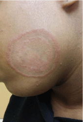
Figure 1
An intensely pruritic 7-cm-diameter annular patch on
the left cheek that has been slowly increasing in size over the last 5 weeks. The ring like lesion appears to be well-demarcated, discrete, hyperpigmented with central clearance and peripheral scaling.
Figure 2
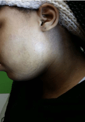
Figure 2
Shows a partially improved lesion with persisted pruritus after 8 weeks of intermittent use of Ciclopirox (0.77%).
Discussion
The patient presented with the typical skin lesions characteristic of tinea incognito and she clearly had a protracted course with multiple ED visits and two failed therapeutic trials. The first trial lasted for 4 weeks using the combination cream which contained in addition to the antifungal ingredient an intermediate potency topical steroid (Betamethasone Dipropionate 0.05% cream) that contributed to this outcome. The second therapeutic trial was the intermittent use of topical Ciclopirox (0.77%) for 8 weeks was not helpful either.
Also, the fact that the patient has been sharing the bed with her pet dog and denied contact with or the presence of similar rash with other household members suggests that the lesion is possibly associated with zoophilic dermatophytes like Microsporum canis which is known to produce more inflammatory response to the lesion than anthropophilic fungi like Trichophytonrubru [2].
The classic tinea corporis lesions are usually annular or circular "ringworm" in shape, well-demarcated, discrete, and with a central clearing surrounded by a raised scaly border. The lesion is usually associated with intense pruritus [1]. If left untreated, the lesion may extend to involve hair follicles, making it more difficult to treat. If the clinician is in doubt of the diagnosis, a confirmation can be reached by visualizing hyphae under microscopic examination of scrapings of the scaly border in a KOH (potassium hydroxide) preparation wet mount [3,4].
The recommended initial treatment is the application of topical antifungal agents like miconazole, clotrimazole, terbinafine, tolnaftate, ketoconazole, or ciclopirox twice daily for a total of 4 weeks. If the initial treatment attempt fails, the patient may need a systemic treatment with either griseofulvin 20 mg/kg/day for 4 to 6 weeks or with terbinafine 250 mg once daily for 2-4 weeks. Another alternative is itraconazole 200 mg daily for one week [3]. Monitoring liver functions is recommended with the use of both itraconazole and terbinafine [1,5].
The patient was treated with terbinafine 250 mg oral tablet daily for 14 Day and was asked to discontinue the ciclopirox cream.
Conclusion
Tinea incognito develops when topical steroids are used and where the clinical appearance as well as the classic features of the initial tinea presentation become distorted as the lesion becomes less scaly, more extensive, more pruritic, and painful. If left untreated, the lesion may extend to involve hair follicles, making it more difficult to treat and it may require the use of systemic anti-fungal agents.
References
- Berger. Committee on Infectious Disease. In: Wolff, Johnson, Suurmond, editors. American Academy of Pediatrics, 2006. Uphold & Graham; 2007.
- Gathings RM, Abide JM, Brodell RT. An unusual inflammatory rash. JAMA Pediatr. 2014; 168: 185-186.
- Elewski BE. Treatment of tinea capitis: beyond griseofulvin. J Am Acad Dermatol. 1999; 40: S27-30.
- De Kock CA, Sampers GH, Knottnerus JA. Diagnosis and management of cases of suspected dermatomycosis in The Netherlands: influence of general practice based potassium hydroxide testing. Br J Gen Pract. 1995; 45: 349-351.
- Brodell RT, Helms SE, Snelson ME. Office dermatologic testing: the KOH preparation. Am Fam Physician. 1991; 43: 2061-2065.