Case Report
Filum Terminale Needle Placement during Caudal Epidural Steroid Injection: A Case Report
Ramsook RR1*, Spinner D1 and Doshi RR2
1Department of Rehabilitation Medicine, Icahn School of Medicine at Mount Sinai, USA
2Division of Pain Medicine, Beth Israel Deaconess Medical Center, USA
*Corresponding author: Ryan R. Ramsook, Department of Rehabilitation Medicine, Icahn School of Medicine at Mount Sinai, USA
Published: 04 Jul, 2016
Cite this article as: Ramsook RR, Spinner D, Doshi RR.
Filum Terminale Needle Placement
during Caudal Epidural Steroid
Injection: A Case Report. Ann Clin Case
Rep. 2016; 1: 1034.
Abstract
Objective: Caudal epidural steroid injections (ESI) are a commonly utilized and efficacious means in which to treat radicular leg symptoms arising from the lumbo-sacral spine. Touhy needles are placed
via fluoroscopic guidance with utilization of live fluoroscopy to ensure epidural administration of
medication. This is a case report of direct needle and catheter placements into and through the filum
terminale during a caudal approach to the epidural space.
Design: Single case report.
Setting: Beth Israel Deaconess Medical Center.
Patient: A 69-year-old woman, who suffered from chronic low back and leg pain from lumbosacral
radiculopathy, failed back surgery syndrome and lumbar facet arthropathy.
Interventions: Caudal epidural steroid injection.
Outcome Measures: Pain reduction.
Results: The needle and catheter placement were confirmed via intrathecal contrast spread to be in
the filum terminale, which resulted in the abortion of the procedure.
Conclusions: This case illustrates a rare incidence of catheter placement directly into the filum
terminale during a routine caudal ESI. Despite a relatively safe and routine intervention, care must
be taken to ensure proper placement of needle, catheter and injectate. While contrast is injected to
ensure appropriate epidural spread, we present, to the best of our knowledge, the first occasion of a
needle being inserted directly into the filum terminale during a caudal ESI.
Keywords: Caudal epidural steroid injection; Filum terminale; Catheter placement, Lumbosacral radiculopathy
Introduction
Therapeutic ESI has been utilized for decades in patients with chronic low back pain particularly lumbar radiculopathy [1]. Caudal ESI have gained significant popularity owing to the ease of the technique and low complication rate [2]. Anatomically, the sacral hiatus, on the dorsum of the S5 vertebrae where it meets the S4 vertebrae, contains the filum terminale. While the thecal sac may vary at its termination site, it typically ends in adults at S2 and in children at S3. This is important for optimum needle placement during caudal ESI and to avoid dural puncture [3]. While the incidence of complications remains low, the most common is insomnia on the night of the injection. Also, transient nonpositional headache and temporary increase in pain at the injection site have been noted [4]. Far more uncommon reactions include vasovagal episodes and local or systemic reactions to either steroid, local anesthetic or contrast media [5]. The filum terminale is a fibrous connective tissue extending from the conus medullaris and attaching to the back of the first segment of the coccyx. As such, this thin structure is an anatomical consideration when performing a caudal ESI. Here we report a previously unrecorded phenomenon of aberrant contrast flow pattern through the filum terminale.
Case Presentation
A 69-year-old woman was scheduled for elective repeat caudal epidural steroid injection at the
Beth Israel Deaconess Medical Center pain management clinic. She presents with a history of L3-4
laminectomy and L4-5 fusion with persistent low back and bilateral lower extremity pain.
The patient stated that her pain control was previously well maintained with her home pain regimen of hydrocodone-acetaminophen and nortriptyline, however recently these medications were rendered ineffective. Two years prior the patient underwent an initial caudal ESI with extended pain relief. More recently her pain progressed and she returned for a repeat injection. Given the good prior response to caudal ESI, she was scheduled for a repeat injection.
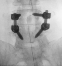
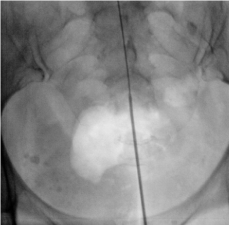
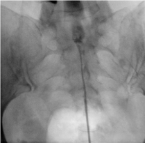
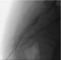
Under AP and lateral fluoroscopic guidance a Coude needle was introduced into the caudal epidural space without advancement beyond S4. An epimed catheter was inserted and tracked to the L4 level (Figure 1 and 2). Contrast was injected and showed thecal spread. The catheter was slowly withdrawn to the S2 level where contrast spread was consistent with thecal spread at each subsequent level. The catheter was then removed and the contrast was inserted through the coude at which point revealed contrast spread through the filum terminale cephalad into the thecal space (Figures 3 and 4).
The thecal space was flushed with preservative free normal saline and the patient began to feel pressure in both legs. At this point the procedure was aborted. The patient was closely monitored post procedure and denied any leg weakness or headaches. This patient with previously strong response to caudal ESI, had a repeat injection with needle placement and catheter advancement into the filum terminale with obvious intrathecal spread.
Figure 1
Figure 2
Figure 3
Figure 3
A-P view of Caudal ESI once catheter was removed with Touhy needle still in place with contrast spread into the thecal space through the filum terminale.
Discussion
Popularity of the caudal ESI remains high secondary to a low complication rate and ease of technique, when performed by a well
trained provider and provided that the patient has minimal anatomic variation. Anatomic considerations are worth reviewing, as proper landmarks can be used to guide optimal needle placement. The dorsally convex sacrum consisting of 5 fused vertebrae is linked to the triangular coccyx consisting of 3 to 5 rudimentary vertebral bones. The sacral hiatus occurs in the midline of the S5 vertebrae dorsally where it meets the S4 vertebrae. The floor of the sacral hiatus is the S5 vertebral body. Within the sacral hiatus are the coccygeal nerve and the filum terminale. In addition to the epidural venous plexus down to S4, there is also epidural fat. The thecal sac can also be found to terminate in this region. While there is some variation in the level of thecal sac termination it can be estimated at S1 in adults and S3 in children [6].
To the best of our knowledge, this is the first case report in the literature of catheter placement into the filum terminale during a caudal ESI. This case highlights a potential anatomical consideration in a commonly utilized procedure. Given how how small the filum terminale is, it is unlikely that a physician would encounter this landmark while performing a caudal ESI. Nonetheless, all physicians should be aware of the possibility of encountering the structure. There is particular significance in practice of this aberrant injection as the filum terminale communicates with the thecal sac. If unrecognized, cannulation of this landmark could cause injectate to be dispersed throughout the thecal sac. This could cause increased complications from the potentially hazardous agents utilized for the injection.
Our patient’s condition required extensive treatment options in
the past. Despite trials of numerous pharmacologic agents along with physical therapy, her pain persisted. The patient had a long standing history of low back pain and she underwent a lumbar laminectomy and fusion 3 years prior. 1 year after her surgery, with pain still being a major issue, she underwent a caudal epidural. The caudal ESI was deemed a success with extended pain relief. Due to the success with her prior caudal ESI, we repeated the procedure to try providing further pain control, In looking at our patient’s contrast flow pattern it is important to note that there is no obvious enhancement of the nerve roots with contrast through the epidural catheter or through the needle alone. Contrast flow could potentially be subdural however no branching was noted making this flow pattern unlikely to be vascular. The contrast flow is highly indicative of filum terminale cannulation.
Figure 4
Conclusion
While the filum terminal is a thin structure it must still be an anatomic consideration when performing a caudal ESI. Our patient had clear needle placement into the filum terminale with contrast spread confirmation. This is, to the best of our knowledge, the first confirmed case of such needle placement. This case highlights the continued need for consideration of various anatomic landmarks during fluoroscopic guided caudal ESI. Physicians should be aware that despite the unlikely possibility of encountering the small filum terminale, injecting through the structure unrecognized may lead to complications via communication with the thecal sac.
References
- Goebert T HW Jr, Jallo SJ, Gardner WJ, Wasmuth CE. Painful radiculopathy treated with epidural injections of procaine and hydrocortisone acetate: results in 113 patients. Anesth Analg. 1961; 40: 130-134.
- Conn A, Buenaventura RM, Datta S, Abdi S, Diwan S. Systematic review of caudal epidural injections in the management of chronic low back pain. Pain Physician. 2009; 12: 109-135.
- Katz J. Caudal approach single injection technique, Katz J editors In: Atlas of Regional Anesthesia. Norwalk, CT, Appleton U Lange, 1994:129.
- Botwin KP, Gruber RD, Bouchlas CG, Torres-Ramos FM, Hanna A, Rittenberg J, et al. Complications of fluoroscopically guided caudal epidural injections. Am J Phys Med Rehabil. 2001; 80: 416-424.
- White AH. Injection techniques for the diagnosis and treatment of low back pain. Orthop Clin North Am. 1983; 14: 553-567.
- Ogoke BA. Caudal epidural steroid injections. Pain Physician. 2000; 3: 305-312.


